Best Keratoconus Specialist in Mumbai — How to Verify ‘Best’ When Your Cornea Is Thinning
Written & medically reviewed by Dr. Jignesh Gala, Vitreoretinal & Cataract Surgeon (FRCS Glasgow), Founder & Director — Crystal Clear Eye Clinic (Andheri West) and Topax Eye Hospital (Vile Parle East), Mumbai. Facts verified: 24 July 2026.
Thank you for reading this post, don't forget to subscribe!⚡ Quick answer: There is no certificate for the best keratoconus specialist in Mumbai — “best” is something you verify. A genuine specialist has cornea-focused training, corneal tomography on-site, disciplined progression tracking, and the full option ladder — cross-linking (C3R/CXL), scleral lenses, Intacs evaluation, honest transplant referral when truly needed. Call 077188 85245 — Dr. Jignesh Gala’s centres in Andheri West and Vile Parle East will walk you through the checklist below.
- Refractive lens exchange rlethe complete guide to clear vision without glasses
- Retina laser treatment in andheri west mumbai prp barrage focal micropulse laser by dr jignesh gala frcs
- Retina surgery in mumbai your complete guide to saving your sight
- Retina treatment in andheri
- Retina treatment in mumbai advanced retinal care by dr jignesh gala
- Rle vs lasik which vision correction surgery is better
- Satisfied patient stories
- Singapore 2021 a year on the founding ophthalmology team of woodlands health
- Smartsurf with customeyes the most advanced glass removal surgery in mumbai and why it outperforms wavelight plus innoveyes
- Surgery packages
- Surgical videos by dr jignesh gala
- Types of intraocular lenses
1. The 8-Point Checklist for a Keratoconus Specialist
Every week, patients from Andheri, Lokhandwala, Versova, Juhu, Bandra and Kandivali arrive holding a Pentacam printout and one frightened question: “Doctor, ab kiske paas jaayein?” The honest answer: not whoever advertises loudest — whoever passes a verifiable checklist. Here are the eight points I would use for my own family:
- Cornea-focused training and volume. A surgeon who occasionally sees a cone between cataract lists differs from one who reads tomography fluently and manages the full spectrum of the disease.
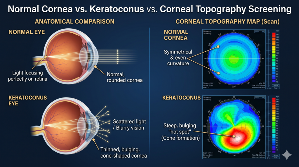
- Corneal tomography on-site. Modern diagnosis runs on Scheimpflug tomography — pachymetry maps, posterior elevation, biomechanical indices (PubMed evidence, AI-tomography evidence). A clinic that sends you elsewhere for the scan cannot own your follow-up data.
- Progression-tracking discipline. Progression is provable from serial tomography — Kmax trends and ABCD grading on the same machine. A specialist who cannot show your own trend line is guessing.
- The full option ladder. CXL (C3R) to halt progression → scleral lenses for vision → Intacs evaluation → and, only for the ~10–15% who truly need it, honest transplant referral. A clinic offering one rung will fit you into that rung.
- Honest outcomes. Cross-linking halts progression; it does not reverse the cone. Intacs improve lens tolerance; they do not stop progression. If someone promises “permanent cure,” walk out.
- Research currency. The evidence moves fast — especially pediatric keratoconus (systematic review, 2024 review). Your doctor should still be reading.
- Language comfort. You will live with this disease for years — you need explanations in Hindi, Marathi, Gujarati or English, not just a printout.
- 7-day access. A sudden hydrops scare or torn RGP lens does not keep Monday-to-Friday hours.
2. Dr. Gala Against the Checklist — Point by Point with Proof
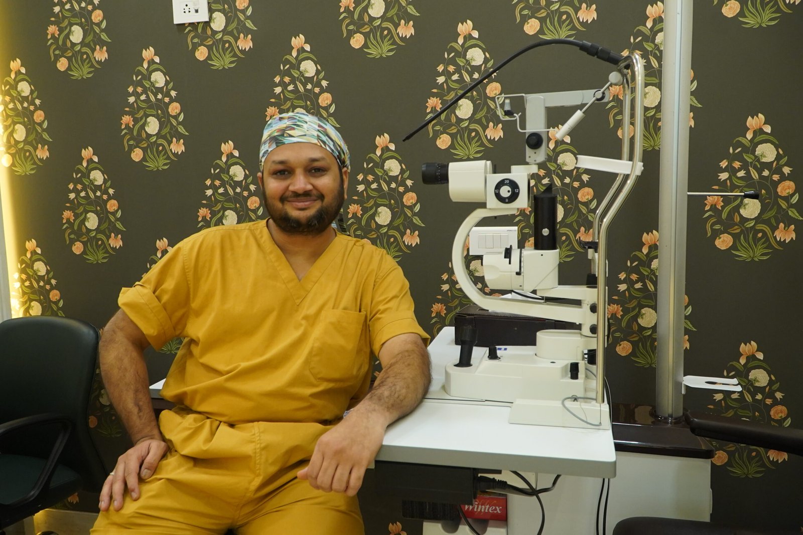
Fair is fair — the same checklist, applied to me:
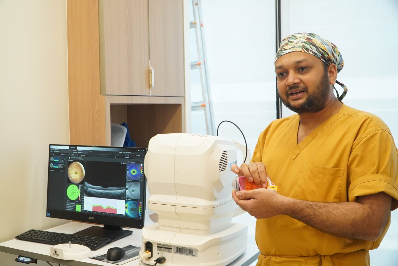
- Training: FRCS (Glasgow), MRCS (Edinburgh), FICO; cornea fellowship at LV Prasad Eye Institute; founding ophthalmology team of Woodlands Health, Singapore; ex-Assistant Professor, B.Y.L. Nair Hospital.
- On-site tomography: corneal topography/tomography at our own centres — no outsourcing, no lost records.
- Progression records: every patient has a serial file — Kmax, thinnest pachymetry, ABCD stage — compared on the same machine.
- Full ladder: CXL/C3R at the centre; scleral-lens fitting coordinated with specialist fitters; Intacs evaluated honestly; the small minority needing DALK/PK counselled truthfully and referred with structured co-management — alternatives exhausted first.
- Honest outcomes: we tell every CXL patient on day one — this stops the worsening; it does not un-thin the cornea.
- Research currency: fully aware of the latest research — unlike old practitioners who stopped reading journals years ago; Cornea, JCRS, Journal of Refractive Surgery, AJO, BJO, IOVS, and six years as a BMJ Case Reports peer reviewer.
- Languages: Hindi, Marathi, Gujarati and English, daily.
- Access: both centres open daily, closing 9 pm, one phone number.
Patients who found us via best eye specialist in Andheri Mumbai or Dr. Jignesh Gala — top eye specialist in Mumbai often arrive after being told “surgery zaroori hai” elsewhere — and leave with a monitoring plan, because their serial scans showed no progression.
3. Understanding Your Diagnosis First
Before choosing a specialist, understand the diagnosis. Keratoconus means the cornea — the eye’s clear front window — gradually thins and bulges forward into a cone. The word sounds alarming; for most patients, the reality is manageable. A “cone” is not a crisis:
- It is common and it is graded. Most cases are mild-to-moderate and slow. Only about 10–15% of patients ever need a corneal transplant — the rest are managed with cross-linking and lenses.
- Progression is measurable, not mystical. Serial tomography (Kmax change, ABCD grading) proves whether your cornea is moving. “Monitoring beats panic” is the evidence-based alternative to operating on every diagnosis.
- Age changes the urgency, not the fear. Pediatric keratoconus progresses faster — children need earlier CXL and tighter follow-up (accelerated CXL evidence, meta-analysis). Our pediatric ophthalmology service in Andheri screens young eye-rubbers aggressively.
- Stop eye rubbing. Whatever your stage, the cheapest intervention is breaking the rubbing habit.
One practical truth: RGP (hard) lenses mould the cornea slightly. For accurate tomography, expect a 2-week RGP washout before your scan — otherwise your “progression” may be mere lens warpage. A clinic that never mentions washout has progression data you should question.
4. Red Flags When Choosing
Three patterns should make you politely collect your file and leave:
- Surgery advised without tomography. If a doctor recommends CXL, Intacs or transplant from a torchlight exam or a single old printout, that is guesswork wearing a white coat. No scan, no surgery decision — period.
- “CXL for everyone.” Cross-linking is for progressive keratoconus. A stable cone unchanged for two years often needs lenses and monitoring, not an operation. The giveaway of a CXL mill: nobody shows you serial scans proving progression.
- No progression records. Ask: “Show me my last two scans side by side.” If the answer involves phoning a scan centre, your trend data does not exist — and every future decision will be made blind.
A confident specialist never fears an informed patient. Browse blogs by Dr. Jignesh Gala — from bakuchiol eye cream and under-eye care to vision correction surgery after 40. Transparency in writing usually means transparency in practice.
5. Why Patients Choose Dr. Jignesh Gala for Keratoconus

Dr. Jignesh Gala owns both Crystal Clear Eye Clinic (Andheri West) and Topax Eye Hospital (Vile Parle East) — an owned centre means the tomography machine, progression file, CXL suite and follow-up calendar answer to one doctor, not a rotating roster of visiting consultants.
✅ Tomography-first decisions: no treatment advised without a current scan on our own machine — RGP washout respected before scanning.
✅ Progression proved, not presumed: serial Kmax and ABCD grading compared visit to visit; CXL only when the trend line says so.
✅ Cross-linking at the centre: C3R/CXL performed in-house, protocol (conventional vs accelerated) matched to age, thickness and cone behaviour — including faster-moving pediatric cones.
✅ Complete ladder, honestly climbed: scleral-lens fitting coordinated with specialist fitters; Intacs evaluation and planning; and for the ~10–15% who genuinely need DALK/PK — truthful counselling, alternatives exhausted first, planned referral with structured co-management. You will never be pushed into an operation you do not need.
✅ Research-current care: fully aware of the latest research — unlike old practitioners who stopped reading journals years ago; Cornea, JCRS, Journal of Refractive Surgery, AJO, BJO and IOVS are his regular reading, plus six years as a BMJ Case Reports peer reviewer.
✅ Whole-eye depth: your keratoconus doctor is also the best retina specialist in Mumbai many families consult — with dedicated services for diabetic retinopathy in Andheri West and recognition as a retina vitreoretinal surgeon.
6. Locations — Keratoconus Care in Andheri West & Vile Parle East
1) Crystal Clear Eye Clinic — Andheri West
📍 Address: Laram Centre CHS, A1-202, Swami Vivekanand Road, above Sunil Jewellers, near NADCO Shopping Centre, Railway Colony, Andheri West, Mumbai 400058 • Open daily, closes 9 pm.
📞 Phone: 077188 85245
💳 Third-party/TPA cashless is done through Crystal Clear Eye Clinic (Andheri West).
Minutes from Lokhandwala, Versova, Juhu, Oshiwara, DN Nagar, Jogeshwari and Seven Bungalows; Bandra, Khar, Santacruz, Goregaon, Malad and Kandivali reach us comfortably. Searching for the best keratoconus specialist in Mumbai from the western suburbs ends here — our page on cashless eye surgery in Mumbai explains the paperwork.

2) Topax Eye Hospital — Vile Parle East
📍 Vile Parle East, Mumbai • 📞 077188 85245
💳 Direct cashless is done through Topax Eye Care (Vile Parle East).
Convenient for Santacruz East, Khar, Bandra East, Kalina, Vakola, Juhu and the airport corridor. As a full hospital, Topax handles everything keratoconus care can lead to — diagnostics, cross-linking, coordination of advanced surgical referrals. Families also use our pages on Contoura LASIK in Mumbai and the Eylea injection price guide — one accountable doctor across conditions.
7. हिंदी • मराठी • ગુજરાતી — Patient Summaries
🇮🇳 हिंदी सारांश — मुंबई में बेस्ट केराटोकोनस स्पेशलिस्ट
बेस्ट केराटोकोनस स्पेशलिस्ट इन मुंबई चुनने का तरीका विज्ञापन नहीं, सत्यापन है: खुद की corneal tomography, हर विज़िट पर Kmax/ABCD से progression tracking, और CXL, scleral lens, Intacs की पूरी सीढ़ी ईमानदारी से। डॉ. जिग्नेश गाला (FRCS ग्लासगो, LVPEI फेलोशिप) अंधेरी वेस्ट और विले पार्ले ईस्ट में यह सब एक छत के नीचे देते हैं। अपॉइंटमेंट: 077188 85245।
🚩 मराठी सारांश — मुंबईतील उत्तम केराटोकोनस तज्ज्ञ
बेस्ट केराटोकोनस स्पेशलिस्ट इन मुंबई शोधताना तीन गोष्टी तपासा: स्वतःची corneal tomography मशीन, Kmax/ABCD नुसार progression ची नोंद, आणि CXL पासून Intacs पर्यंत सर्व पर्याय प्रामाणिकपणे सांगणारा डॉक्टर. डॉ. जिग्नेश गाला (FRCS ग्लासगो, LVPEI फेलोशिप) अंधेरी वेस्ट व विले पार्ले ईस्ट येथे तपासणी, क्रॉस-लिंकिंग व फॉलो-अप एकाच ठिकाणी करतात. भेटीसाठी: 077188 85245.
🟡 ગુજરાતી સારાંશ — મુંબઈમાં બેસ્ટ કેરાટોકોનસ સ્પેશિયાલિસ્ટ
બેસ્ટ કેરાટોકોનસ સ્પેશિયાલિસ્ટ ઇન મુંબઈ પસંદ કરતાં પુરાવા જુઓ: પોતાની corneal tomography છે કે નહીં, Kmax/ABCD થી progression ટ્રૅક થાય છે કે નહીં, અને CXL, scleral lens, Intacs — બધા વિકલ્પો પ્રમાણિકતાથી સમજાવાય છે કે નહીં. ડૉ. જિગ્નેશ ગાલા (FRCS ગ્લાસગો, LVPEI ફેલોશિપ) અંધેરી વેસ્ટ અને વિલે પાર્લે ઈસ્ટમાં આખી સારવાર એક જ જગ્યાએ આપે છે. એપોઈન્ટમેન્ટ: 077188 85245.
8. ❓ 8 Frequently Asked Questions (English + Hinglish)
1. Best keratoconus specialist in Mumbai kaise choose karein?
Checklist se verify karein: cornea-focused training, clinic mein khud ki corneal tomography, serial scans se progression tracking (Kmax/ABCD), CXL se lenses aur Intacs tak ka full option ladder, aur honest outcomes. Jo doctor scan ke bina surgery bol de, usse door rahein. Dr. Gala ke centres par yahi checklist test kar sakte hain — call 077188 85245.
2. What qualifications should a keratoconus specialist have?
Recognized surgical fellowships (FRCS/MRCS/FICO level), cornea or anterior-segment fellowship experience (LVPEI-type institutes are the Indian benchmark), active journal reading, and — practically — on-site tomography plus a documented progression-tracking system. Degrees get a doctor into the room; discipline with your serial scans protects your cornea.
3. Keratoconus consultation fees in Mumbai kitni hai?
Consultation typically ₹800–2,000 across Mumbai; corneal tomography/Pentacam scan ₹1,200–2,500 (advanced tomography up to ₹5,000) — verified 24 July 2026. Exact quote ke liye call karein 077188 85245 — number milega, runaround nahi.
4. Kya keratoconus ka ilaaj possible hai?
Haan — lekin sahi matlab samajhiye. Cross-linking (C3R/CXL) progression rok deta hai, cone reverse nahi karta. Vision scleral lenses se bahut achhi ho jaati hai; Intacs kuch patients ki lens tolerance improve karte hain. Sirf 10–15% patients ko kabhi transplant chahiye hota hai. Sahi specialist ke saath zyadatar patients normal life jeete hain.
5. Do I need tomography before any treatment decision?
Yes — absolutely. Corneal tomography (Scheimpflug imaging) is the objective evidence of where your cone is and whether it is moving. No responsible specialist advises CXL, Intacs or transplant without it. RGP wearers need a 2-week washout before scanning.
6. How often should keratoconus be monitored?
Every 6–12 months with repeat tomography on the same machine — every 3–6 months for children and teens (pediatric keratoconus progresses faster) and after any treatment change. One scan is a photograph; serial scans are a diagnosis.
7. Best keratoconus specialist kahan milein Mumbai mein?
Western suburbs ke liye: Crystal Clear Eye Clinic — Laram Centre CHS, A1-202, S.V. Road, above Sunil Jewellers, near NADCO Shopping Centre, Andheri West 400058; aur Topax Eye Hospital, Vile Parle East. Phone: 077188 85245. Open daily, closes 9 pm. Lokhandwala, Versova, Juhu, Oshiwara, Bandra, Khar, Santacruz — sab se aasaan access.
8. Can I get a second opinion for keratoconus from Dr. Gala?
Yes — actively encouraged. Bring your old scans and prescriptions; Dr. Gala will do fresh tomography, compare where possible, and tell you plainly whether the advised treatment is warranted. Many “surgery zaroori hai” cases need only monitoring and lenses. Call 077188 85245 to book.
13. 📖 Related Reading — Complete Eye-Care Library by Dr. Jignesh Gala (78 guides)
Every guide below is written by the same surgeon who would operate on you:
From drjigneshgalaeye.com
- Drjigneshgalaeye.com
- A year at l v prasad eye institute how my vitreoretinal fellowship shaped the surgeon i am today
- About us
- Anti vegf injection cost in mumbai 2026 every drug every price explained honestly by a retina specialist
- B y l nair hospital 2019 2020 my sixteen months as assistant professor of ophthalmology
- Bakuchiol eye cream a gentle revolution in under eye care
- Best eye doctor for retina in mumbai dr jignesh gala retina vitreoretinal surgeon
- Best eye specialist in andheri mumbai
- Best retina specialist mumbai dr jignesh gala
- Best vision correction surgery after 40 what are your options
- Blogs by dr jignesh gala
- Cashless eye surgery in mumbai dr jignesh gala
- Diabetic retinopathy specialist in andheri west mumbai advanced screening injections laser vitrectomy by dr jignesh gala frcs
- Discover crystal clear vision with contoura lasik in mumbai
- Dr jignesh gala top eye specialist in mumbai
- Drjignesh gala premier pediatric ophthalmologistandheri mumbai
- Eylea injection in mumbai cost uses side effects the 2026 price revolution explained by a retina specialist
- Glaucoma treatment in andheri mumbai
- How to prevent dry eyes in winter
- Hvf test in andheri west mumbai visual field test cost procedure how to read your report by a specialist
- I see a curtain or shadow in my vision
- Inside my six years as a bmj case reports peer reviewer
- Inside the fico 2016 the only worldwide medical specialty examination
- Inside the frcs ophth glasgow how a 425 year old royal college tests your surgical knowledge
- Intravitreal injection in andheri west mumbai painless anti vegf eye injections by dr jignesh gala frcs
- Know your eye doctor
- Laser cataract surgery in andheri mumbai dr jignesh gala
- Laser cataract surgery
- Lasik eye surgery in andheri mumbai dr jignesh gala
- Lasik eye surgery in bandra mumbai dr jignesh gala
- Lasik eye surgery in juhu dr jignesh gala
- Lasik eye surgery in lokhandwala dr jignesh gala
- Lasik eye surgery in oshiwara dr jignesh gala
- Lasik in santacruz mumbai
- Lasik in versova mumbai dr jignesh gala
- Lasik surgery in andheri mumbai
- Most common types of refractive eye surgery which one is right for you
- Mrcs edinburgh how i joined a royal college founded in 1505
- Multifocal trifocal iol guide complete patient guide
- Oct eye test in andheri west mumbai cost procedure report why the specialist reading it matters
- People do ask google about this eye issue
- Refraction in real time how the royal college of ophthalmologists tested me in kuching 2017
- Refractive lens exchange rlethe complete guide to clear vision without glasses
- Retina laser treatment in andheri west mumbai prp barrage focal micropulse laser by dr jignesh gala frcs
- Retina surgery in mumbai your complete guide to saving your sight
- Retina treatment in andheri
- Retina treatment in mumbai advanced retinal care by dr jignesh gala
- Rle vs lasik which vision correction surgery is better
- Satisfied patient stories
- Singapore 2021 a year on the founding ophthalmology team of woodlands health
- Smartsurf with customeyes the most advanced glass removal surgery in mumbai and why it outperforms wavelight plus innoveyes
- Surgery packages
- Surgical videos by dr jignesh gala
- Types of intraocular lenses
- Types of refractive surgeries
- Types of retina surgeries
- Understanding and preventing digital eye strain
From crystalcleareye.in
- Crystal Clear Eye Clinic — Home
- About Crystal Clear Eye Clinic, Andheri West
- Eye Health Blog — Crystal Clear Eye Clinic
- Contact & Directions — Andheri West
- Glaucoma Treatment in Andheri, Mumbai
- LASIK Surgery in Andheri, Mumbai
- Premium IOL Selection Guide 2026
- Laser Cataract Surgery in Andheri, Mumbai
- Best Eye Specialist in Andheri, Mumbai
- Best Retina Specialist in Mumbai
- Best Vitrectomy Surgery in Mumbai — Expert Retinal Care
- Diabetic Eye Care in Mumbai
- Diabetic Retinopathy in Andheri
- Diabetic Macular Edema (DME) Treatment in Mumbai
- Macular Edema Treatment in Andheri West
- ARMD Treatment in Andheri West, Mumbai
- Retina Treatment in Andheri
- Vitreoretinal Treatment in Andheri
- Cashless Eye Surgery in Mumbai
- Sudden Floaters? Retina Check-up in Mumbai
- What Is an Intravitreal Eye Injection?
Your retina deserves a specialist. Today, not someday.
Keratoconus is a 20-year relationship, not a one-visit illness — choose the specialist whose checklist survives your questions.
📍 Andheri West: Laram Centre CHS, A1-202, S.V. Road, above Sunil Jewellers, near NADCO Shopping Centre, Mumbai 400058
📍 Vile Parle East: Topax Eye Hospital • Open daily, closes 9 pm
Dr. Jignesh Gala, FRCS (Glasgow) — Vitreoretinal & Cataract Surgeon • drjigneshgalaeye.com • crystalcleareye.in
Medical disclaimer: This page is educational and does not replace a personal consultation. Prices are indicative, verified on 24 July 2026, and may change. Every eye is different — decisions are made only after examination by Dr. Jignesh Gala.
Research & Evidence Library — 108 Verified References
Verified live in July 2026 via official NCBI E-utilities — including research from Cornea, JCRS, Journal of Refractive Surgery, AJO, BJO, IOVS, Eye & Contact Lens, IJO, Acta Ophthalmologica, Graefe’s Archive and TVST. The same journals Dr. Gala reads. Click any title to check the science yourself.
KC: Corneal Cross-Linking: Long-Term Outcomes
- Long-term outcomes of corneal crosslinking. — Current opinion in ophthalmology, 2024
- Long term results of accelerated corneal collagen cross-linking in pediatric keratoconus. — European journal of ophthalmology, 2021
- Long-Term Outcomes After Corneal Cross-linking for Progressive Keratoconus and Corneal Ectasia: A 10-Year Follow-Up of the Pivotal Study. — Eye & contact lens, 2023
- Corneal Collagen Cross-Linking for Keratoconus in Pediatric Patients-Long-Term Results. — Cornea, 2017
- Short- and long-term safety and efficacy of corneal collagen cross-linking in progressive keratoconus: A systematic review and meta-analysis of randomized controlled trials. — Taiwan journal of ophthalmology, 2023
- Corneal Allogenic Intrastromal Ring Segments (CAIRS) Combined With Corneal Cross-linking for Keratoconus. — Journal of refractive surgery (Thorofare, N.J. : 1995), 2018
- Safety and Efficacy of Corneal Cross-Linking in Patients Affected by Keratoconus: Long-Term Results. — Medical sciences (Basel, Switzerland), 2023
- Long-term changes in crystalline lens transparency after accelerated transepithelial corneal cross-linking in patients with keratoconus. — European journal of ophthalmology, 2023
- Long-term Outcomes of Accelerated Corneal Cross-linking in the Treatment of Keratoconus: Comparison of Hypotonic Riboflavin Solution With Standard Riboflavin Solution. — Journal of refractive surgery (Thorofare, N.J. : 1995), 2020
- Long-term Evaluation of Corneal Biomechanical Properties After Corneal Cross-linking for Keratoconus: A 4-Year Longitudinal Study. — Journal of refractive surgery (Thorofare, N.J. : 1995), 2018
- Cutting-edge cross-linking biomaterials advancing ophthalmic therapeutics. — Progress in retinal and eye research, 2026
- Five-year corneal cross-linking outcomes: A Save Sight Keratoconus Registry Study. — Clinical & experimental ophthalmology, 2023
KC: Corneal Tomography / Pentacam Diagnosis & Progression
- Corneoscleral Morphology in Keratoconus and Its Association With Corneal Tomography and Biomechanics. — Translational vision science & technology, 2026
- Simultaneous Topography-guided Custom Ablation With Corneal Cross-linking for Keratoconus: 10-Year Prospective Outcomes. — Journal of refractive surgery (Thorofare, N.J. : 1995), 2023
- Superiority of Baseline Biomechanical Properties over Corneal Tomography in Predicting Keratoconus Progression. — Turkish journal of ophthalmology, 2021
- Machine Learning-Based Identification of Risk Factors of Keratoconus Progression Using Raw Corneal Tomography Data. — Cornea, 2024
- Topical Losartan for Treating Corneal Haze After Ultraviolet-A/Riboflavin Collagen Cross-Linking. — Cornea, 2024
- Repeatability and Comparison of Corneal Tomography in Mild to Severe Keratoconus Between the Anterior Segment OCT MS-39 and Pentacam HR. — Journal of refractive surgery (Thorofare, N.J. : 1995), 2022
- Keratoconus tomographic indices in osteogenesis imperfecta. — Graefe’s archive for clinical and experimental ophthalmology = Albrecht von Graefes Archiv fur klinische und experimentelle Ophthalmologie, 2023
- Belin ABCD Progression Display Identifies Keratoconus Progression Earlier Than Conventional Metrics. — American journal of ophthalmology, 2022
- Scheimpflug-Derived Keratometric, Pachymetric and Pachymetric Progression Indices in the Diagnosis of Keratoconus: A Systematic Review and Meta-Analysis. — Clinical ophthalmology (Auckland, N.Z.), 2023
- Corneal cross-linking in patients with keratoconus: up to 13 years of follow-up. — Graefe’s archive for clinical and experimental ophthalmology = Albrecht von Graefes Archiv fur klinische und experimentelle Ophthalmologie, 2023
- Influence of Keratoconus Severity on Detecting True Progression with Scheimpflug Imaging and Anterior Segment Optical Coherence Tomography. — Life (Basel, Switzerland), 2023
- Artificial intelligence for detecting keratoconus. — The Cochrane database of systematic reviews, 2023
KC: Artificial Intelligence Detection (Tomography/Biomechanics)
- Enhancing Early Keratoconus Detection With Multimodal Machine Learning: Integrating Tomography, Biomechanics, and Clinical Risk Factors. — American journal of ophthalmology, 2025
- Keratoconus: imaging modalities and management. — Medical hypothesis, discovery & innovation ophthalmology journal, 2024
- [Detection of Subclinical Keratoconus]. — Klinische Monatsblatter fur Augenheilkunde, 2022
- Advancing subclinical keratoconus detection using polarization-sensitive optical coherence tomography and artificial intelligence. — Biophotonics discovery, 2026
- Optimized Artificial Intelligence for Enhanced Ectasia Detection Using Scheimpflug-Based Corneal Tomography and Biomechanical Data. — American journal of ophthalmology, 2023
- Artificial Intelligence-Derived Biomechanical Index for Early Corneal Ectasia Detection: Advancing Beyond Tomography. — Ophthalmology science, 2026
- Early Detection of Keratoconus: Diagnostic Advances and Their Impact on Visual Outcomes: A Systematic Review. — Medicina (Kaunas, Lithuania), 2025
- Artificial intelligence in ophthalmology: opportunities, challenges, and ethical considerations. — Medical hypothesis, discovery & innovation ophthalmology journal, 2025
- A Novel Chaos-inspired Artificial Intelligence-based Model for Keratoconus Prediction. — Journal of ophthalmic & vision research, 2026
- Risk factors for postlaser refractive surgery corneal ectasia. — Current opinion in ophthalmology, 2020
- Machine Learning Algorithms to Detect Subclinical Keratoconus: Systematic Review. — JMIR medical informatics, 2021
- Multimodal diagnostics for keratoconus and ectatic corneal diseases: a paradigm shift. — Eye and vision (London, England), 2023
KC: Genetics, Pathophysiology & Biomechanics
- Advances in keratoconus animal models: From genetics to biomechanics. — Experimental eye research, 2025
- A review of keratoconus: Diagnosis, pathophysiology, and genetics. — Survey of ophthalmology, 2017
- [KERATOCONUS: DIAGNOSIS AND INNOVATIONS]. — Harefuah, 2025
- The pathogenesis of keratoconus. — Eye (London, England), 2014
- Nutritional and Metabolic Imbalance in Keratoconus. — Nutrients, 2022
- Update on the Physiopathology of Keratoconus. — Medical sciences (Basel, Switzerland), 2026
- The Proteins of Keratoconus: a Literature Review Exploring Their Contribution to the Pathophysiology of the Disease. — Advances in therapy, 2019
- Experimental models for keratoconus: Insights and challenges. — Experimental eye research, 2024
- Tear Proteomic Analysis From Offspring of Keratoconus Patients: New Insights Into Corneal Biomechanical Weakness and Disease Risk Stages. — Investigative ophthalmology & visual science, 2025
- Biomechanical and inflammatory pathways underlying the genetic architecture of keratoconus: A genomic SEM study. — Experimental eye research, 2026
- The importance of corneal biomechanics in assessing first degree family members of keratoconus patients. — Romanian journal of ophthalmology, 2018
- Keratoconus Prevalence in Families: A French Study. — Cornea, 2020
KC: Accelerated Vs Conventional Cross-Linking (Epi-On)
- Keratoconus Treatment Algorithm. — Ophthalmology and therapy, 2017
- Expanding indications for corneal cross-linking. — Current opinion in ophthalmology, 2023
- Accelerated Epi-On Versus Standard Epi-Off Corneal Collagen Cross-Linking for Progressive Keratoconus in Pediatric Patients: Five Years of Follow-Up. — Cornea, 2020
- Accelerated Epi-On Versus Standard Epi-Off Corneal Collagen Cross-Linking for Progressive Keratoconus in Pediatric Patients. — Cornea, 2017
- A Transepithelial Corneal Cross-linking (CXL) Protocol Providing the Same Biomechanical Strengthening as Accelerated Epithelium-off CXL. — Journal of refractive surgery (Thorofare, N.J. : 1995), 2025
- A Review of Keratoconus Cross-Linking Treatment Methods. — Journal of clinical medicine, 2025
- Accelerated epithelium-on or accelerated epithelium-off corneal collagen cross-linking: Contralateral comparison study. — Taiwan journal of ophthalmology, 2020
- Progressive high-fluence epithelium-on accelerated corneal crosslinking: a novel corneal photodynamic therapy for early progressive keratoconus. — Frontiers in medicine, 2023
- Efficacy and safety of progressive fluence pulsed light epithelium-on accelerated corneal cross-linking for progressive keratoconus: 18-month prospective results. — International journal of ophthalmology, 2026
- Visual and Topographic Improvement with Epithelium-On, Oxygen-Supplemented, Customized Corneal Cross-Linking for Progressive Keratoconus. — Journal of clinical medicine, 2020
- Optimization of Oxygen Dynamics, UV-A Delivery, and Drug Formulation for Accelerated Epi-On Corneal Crosslinking. — Current eye research, 2020
- Epithelium-on Corneal Cross-linking Using a Modified Penetration Enhancer Without Supplemental Oxygen or Iontophoresis. — Journal of refractive surgery (Thorofare, N.J. : 1995), 2026
- Topo-Pachimetric Accelerated Epi-On Cross-Linking Compared to the Dresden Protocol Using Riboflavin with Vitamin E TPGS: Results of a 2-Year Randomized Study. — Journal of clinical medicine, 2021
- Management of Tomographically Suspicious Fellow Eyes of Young Patients With Unilateral Clinically Evident Keratoconus: Accelerated Epithelium-On Corneal Crosslinking Versus Observation. — Cornea, 2025
- Sterile corneal infiltrates after corneal collagen cross-linking: evaluation of risk factors. — Acta ophthalmologica, 2017
KC: Scleral Lenses: Visual Rehabilitation
- Keratoconus Diagnosis and Treatment: Recent Advances and Future Directions. — Clinical ophthalmology (Auckland, N.Z.), 2023
- Visual rehabilitation with mini scleral contact lenses in scarred corneas. — Journal francais d’ophtalmologie, 2024
- Efficacy of mini-scleral lenses in visual rehabilitation for corneal disorders: a prospective study. — BMC ophthalmology, 2025
- Visual rehabilitation using SPOT(®) scleral lenses in patients with keratoconus. — European journal of ophthalmology, 2026
- Visual rehabilitation using mini-scleral contact lenses after penetrating keratoplasty. — Arquivos brasileiros de oftalmologia, 2017
- Refractive approaches to visual rehabilitation in patients with keratoconus. — Current opinion in ophthalmology, 2020
- Visual Rehabilitation of Bilateral Keratoconus With Customized Scleral Lenses: A Case Report. — Cureus, 2026
- Safety and Efficacy of Scleral Lenses for Keratoconus. — Optometry and vision science : official publication of the American Academy of Optometry, 2020
- Contact lenses for visual rehabilitation in post-keratoplasty eyes: A review. — Indian journal of ophthalmology, 2025
- Scleral contact lenses for visual rehabilitation after penetrating keratoplasty: long term outcomes. — Contact lens & anterior eye : the journal of the British Contact Lens Association, 2014
- Mini-Scleral Lens: An Effective Mode of Visual Rehabilitation in Eyes With Resolved Hydrops. — Eye & contact lens, 2024
- Influence of Scleral Contact Lenses on Optical Coherence Tomography Parameters in Keratoconus Patients. — Diagnostics (Basel, Switzerland), 2025
- Impact of Scleral Lenses on Visual Acuity and Ocular Aberrations in Corneal Ectasia: A Comprehensive Review. — Journal of personalized medicine, 2024
- Irregular Corneas: Improve Visual Function With Scleral Contact Lenses. — Eye & contact lens, 2018
- Visual performance of scleral lenses and their impact on quality of life in patients with irregular corneas. — Arquivos brasileiros de oftalmologia, 2018
KC: Intracorneal Ring Segments (Intacs)
- Intracorneal Ring Segments in Keratoconus: A Narrative Literature Review. — Clinical ophthalmology (Auckland, N.Z.), 2025
- Intracorneal ring segment implantation in advanced Keratoconus. — European journal of ophthalmology, 2023
- Clinical Outcomes and Complications After INTACS Implantation in Keratoconus: A Systematic Review. — Journal of clinical medicine, 2026
- Refractive and aberrometric outcomes of intracorneal ring segments for keratoconus: mechanical versus femtosecond-assisted procedures. — Ophthalmology, 2009
- [Intacsintracorneal ring segments in keratoconus]. — Der Ophthalmologe : Zeitschrift der Deutschen Ophthalmologischen Gesellschaft, 2013
- Outcomes of Single Segment Implantation of Conventional Intacs versus Intacs SK for Keratoconus. — Journal of ophthalmic & vision research, 2014
- Implantation of intracorneal ring segments in pediatric patients: long-term follow-up. — International medical case reports journal, 2018
- Intracorneal ring segments for keratoconus correction: long-term follow-up. — Journal of cataract and refractive surgery, 2006
- Intracorneal ring segments in ectatic corneal disease – a review. — Clinical & experimental ophthalmology, 2010
- Intracorneal Ring Segments Followed by Simultaneous Topography-Guided Removal of Epithelium and Stroma With Accelerated Collagen Cross-Linking For Keratoconus (I-TRESK/CXL). — Asia-Pacific journal of ophthalmology (Philadelphia, Pa.), 2020
- Intacs in advanced keratoconus. — Cornea, 2008
- Intracorneal ring segment implantation for the management of keratoconus: safety and efficacy. — Journal of cataract and refractive surgery, 2007
- Corneal crosslinking and intracorneal ring segments for keratoconus: A randomized study of concurrent versus sequential surgery. — Journal of cataract and refractive surgery, 2019
- Long-term follow up of intracorneal ring segment implantation in 932 keratoconus eyes. — Journal francais d’ophtalmologie, 2020
- Modified intracorneal ring segment implantations (INTACS) for the management of moderate to advanced keratoconus: efficacy and complications. — Cornea, 2006
KC: Pediatric Keratoconus Cross-Linking
- Progression of Pediatric Keratoconus After Corneal Cross-Linking: A Systematic Review and Pooled Analysis. — Cornea, 2022
- Corneal Cross-Linking for Pediatric Keratoconus. — Diagnostics (Basel, Switzerland), 2024
- Accelerated Corneal Collagen Cross-Linking in Pediatric Keratoconus. — Journal of current ophthalmology, 2021
- Corneal collagen cross-linking in pediatric keratoconus with three protocols: a systematic review and meta-analysis. — Journal of AAPOS : the official publication of the American Association for Pediatric Ophthalmology and Strabismus, 2020
- Accelerated Corneal Cross-linking in Pediatric Patients with Progressive Keratoconus: 12-Month Follow-up Results. — Nigerian journal of clinical practice, 2024
- Investigation of Epithelial Remodeling After Accelerated Epi-Off Cross-linking in Pediatric Keratoconus. — Eye & contact lens, 2025
- Corneal Cross-Linking as Treatment in Pediatric Keratoconus: Comparison of Two Protocols. — Journal of ophthalmology, 2021
- Corneal Collagen Cross-Linking for Progressive Keratoconus in Pediatric Patients: Up to 14 Years of Follow-up. — American journal of ophthalmology, 2023
- Astigmatic Vectorial Analysis in Pediatric Keratoconus After Unilateral Cross-Linking Treatment. — Cornea, 2022
- Corneal Cross-Linking for Pediatric Keratcoconus Review. — Cornea, 2018
- Outcomes of accelerated corneal cross-linking for pediatric and adult keratoconus: a comparative study. — International ophthalmology, 2024
- Corneal Cross-Linking for Paediatric Keratoconus: A Systematic Review and Meta-Analysis. — Journal of clinical medicine, 2021
- Comparison of Two Different Accelerated Corneal Cross-linking Procedure Outcomes in Patients with Keratoconus. — Balkan medical journal, 2020
- Bilateral simultaneous accelerated corneal collagen cross-linking in pediatric keratoconus: 24-month follow-up results. — Indian journal of ophthalmology, 2025
- Safety and Efficacy of Corneal Cross-linking in Pediatric Patients with Keratoconus and Vernal Keratoconjunctivitis. — Middle East African journal of ophthalmology, 2019