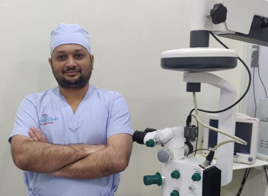
Complicated Eye Surgeries in Andheri West, Mumbai — When Every Other Door Closes, This Is Where Complex Cases Come
Written & performed by Dr. Jignesh Gala, Vitreoretinal & Cataract Surgeon (FRCS Glasgow), Founder & Director — Crystal Clear Eye Clinic (Andheri West) and Topax Eye Hospital (Vile Parle East), Mumbai. Facts verified: 24 July 2026.
Thank you for reading this post, don't forget to subscribe!🚨 EYE EMERGENCY? Sudden shower of floaters, flashes of light, or a curtain/shadow falling across your vision = possible retinal detachment. This is the one eye condition where hours matter. Call 077188 85245 now — retinal emergencies are seen same-day at Crystal Clear Eye Clinic, Andheri West and Topax Eye Hospital, Vile Parle East. Open daily, closes 9 pm.
⚡ Quick answer: A “complicated” eye surgery is one where anatomy, scarring, diabetes, trauma — or a previous failed surgery — has made the standard operation impossible or dangerous. These cases need a vitreoretinal surgeon: retinal detachment with PVR, diabetic tractional detachment, macular hole, dropped nucleus, dislocated lens, scleral-fixated IOL, endophthalmitis, open-globe trauma. In Andheri West, Mumbai, Dr. Jignesh Gala (FRCS Glasgow) performs the full range of complicated retina and cataract surgeries at his own centres — Crystal Clear Eye Clinic and Topax Eye Hospital — including cases referred after failed surgery elsewhere. Complicated eye surgery cost in Mumbai ranges from ₹40,000 (simple vitrectomy) to ₹1,50,000 (complex diabetic/detachment). Call 077188 85245.
1. What Makes an Eye Surgery “Complicated”?
Most eye surgery is routine — a 10-minute cataract removal, a straightforward injection. But some eyes arrive with the rulebook already torn up. A surgery becomes complicated when:
- Tissue is scarred or pulling — diabetic membranes strangling the retina, PVR (proliferative vitreoretinopathy) shortening a detached retina like a tightened drawstring;
- Anatomy has collapsed — a lens nucleus dropped into the vitreous cavity, an IOL floating where no lens should be, a globe ruptured by trauma;
- Infection is racing — endophthalmitis can destroy an eye in 48–72 hours;
- The first surgery failed — recurrent detachment, persistent macular hole, a cataract surgery that ended in complication elsewhere;
- Multiple structures need repair at once — combined cornea-lens-retina injuries that no single-specialist surgeon can finish alone.
These are the cases where the operating surgeon’s training decides the outcome. Dr. Jignesh Gala is a vitreoretinal surgeon — fellowship-trained at L.V. Prasad Eye Institute, one of Asia’s finest retina institutes, with an FRCS (Glasgow), MRCS (Edinburgh), the international FICO qualification, a year on the founding ophthalmology team of Woodlands Health, Singapore, and sixteen months as Assistant Professor of Ophthalmology at B.Y.L. Nair Hospital, Mumbai — where complicated cases are the daily bread. He owns and operates at both his centres — Crystal Clear Eye Clinic, Andheri West and Topax Eye Hospital, Vile Parle East — so a complicated case never gets referred away mid-treatment. Patients searching for a complicated eye surgery in Mumbai, a complex retina surgeon in Andheri West, or the best eye specialist in Mumbai for a difficult case — this page is your honest, detailed map of what each surgery involves, what it costs, and what outcomes to expect.
🏅 Credentials strip: FRCS (Glasgow) • MRCS (Edinburgh) • FICO (International Council of Ophthalmology) • Vitreoretinal Fellowship, L.V. Prasad Eye Institute • Former Assistant Professor, B.Y.L. Nair Hospital • Founding ophthalmology team, Woodlands Health Singapore • Six years as a BMJ Case Reports peer reviewer — the journal trusts him to judge other doctors’ complex cases. Fully aware of the latest research — unlike old practitioners who stopped reading journals years ago — with 120+ recent PubMed references backing every technique on this page.
2. The 10 Complicated Surgeries — Part 1: Retina
Each complex eye surgery card below follows the same honest format: what the condition is, when it becomes complicated, what Dr. Gala actually does in theatre, and what outcomes the research supports.
🔴 Surgery 1: Retinal Detachment Repair (Vitrectomy • Buckle • Pneumatic Retinopexy)
What it is: the retina — the eye’s photographic film — peels away from its blood supply. Untreated, it blinds. Every hour counts once the macula is threatened.
When it becomes complicated: long-standing detachments, PVR scarring, giant tears, detachments in highly myopic eyes, or re-detachment after previous surgery.
What Dr. Gala does: chooses the right weapon per eye — 23/25-gauge micro-incision vitrectomy with gas or silicone oil tamponade, scleral buckling for younger/phakic patients, or office-based pneumatic retinopexy for selected superior detachments. The PIVOT trial-era evidence guides this choice (pneumatic vs vitrectomy evidence), and modern reviews confirm ~90% single-surgery success in expert hands (2024 review).
Honest outcomes: 90–95% reattached with one surgery; final vision depends on whether the macula was still attached when you arrived — which is why the emergency banner tops this page.
🟣 Surgery 2: Macular Hole Surgery (ILM Peel + Inverted Flap)
What it is: a full-thickness gap in the macula — the central reading spot. Vision distorts, then drops; holes never close on their own.
When it becomes complicated: large holes (>400 µm), chronic holes, holes in high myopes, and holes that failed to close after a first surgery elsewhere.
What Dr. Gala does: vitrectomy with internal limiting membrane (ILM) peeling, using the inverted flap technique for large or stubborn holes — the approach with the strongest closure evidence (ILM peel vs inverted flap evidence), with 2026 refinements like free-flap covering already on his reading list (2026 technique). Gas tamponade + guided positioning after.
Honest outcomes: 90–95% closure rates; large/chronic holes 85–90% with flap techniques. Vision improves over 3–6 months.
🟡 Surgery 3: Epiretinal Membrane (ERM) Peeling
What it is: a cellophane-like scar wrinkling the macula — words bend, faces warp, straight lines kink.
When it becomes complicated: thick membranes stuck to vessels, membranes after previous retinal surgery or vein occlusion, and lamellar holes masquerading as simple ERMs.
What Dr. Gala does: micro-incision vitrectomy with careful membrane + ILM peeling under stain — a surgery of millimetres and patience, timed when distortion (not just vision numbers) justifies it — related: our macular edema treatment guide.
Honest outcomes: most patients gain 2+ lines and major distortion relief; improvement builds over months. Surgery too early = unnecessary risk; too late = limited gains. Timing is the skill.
🟠 Surgery 4: Diabetic Vitrectomy (Tractional Detachment • Vitreous Hemorrhage)
What it is: end-stage diabetic retinopathy — blood filling the eye (vitreous hemorrhage) or scar tissue dragging the retina off (tractional detachment).
When it becomes complicated: always. These are among the hardest surgeries in ophthalmology — fragile ischemic retina, active neovascularization, bleeding mid-surgery, and often both eyes at risk.
What Dr. Gala does: pre-operative anti-VEGF injection to quieten vessels, then meticulous membrane delamination/segmentation with 25-gauge vitrectomy, endolaser and tamponade as needed — sequenced with PRP laser strategy per the latest randomized evidence (PRP vs anti-VEGF evidence). Modern series report anatomical success above 85–90% even in tractional detachments (diabetic TRD outcomes).
Honest outcomes: anatomy restored in the large majority; visual recovery is limited by how long the macula was detached and how damaged the diabetic retina already is. Sugar control afterwards is half the surgery.
🔵 Surgery 5: Complex Cataract Surgery (Subluxated Lens • Posterior Polar • Small Pupil • Rock-Hard)
What it is: cataract surgery — but in an eye where the routine playbook fails: the lens hangs by loose threads (subluxation/pseudoexfoliation), the capsule hides a posterior polar landmine, the pupil won’t dilate, or the lens is too hard for standard phaco.
When it becomes complicated: pseudoexfoliation with weak zonules (evidence), posterior polar cataracts (capsule rupture risk up to 26% in average hands), traumatic subluxation, iris scarring (evidence), white/brown mature cataracts.
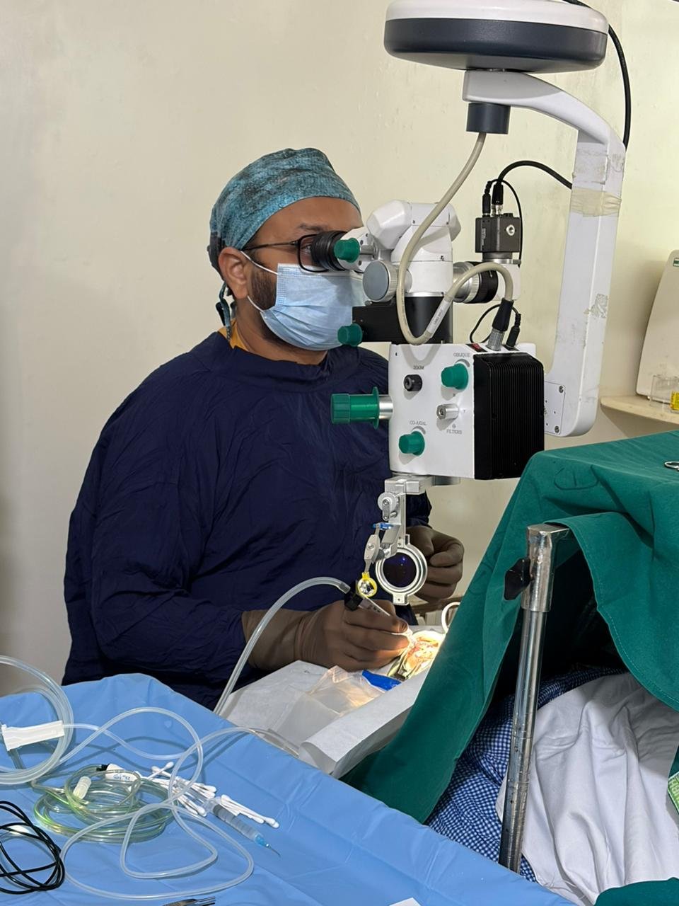
What Dr. Gala does: as a surgeon who operates both the front and back of the eye, he brings retina-grade backup to cataract surgery: capsular support devices, iris hooks, gentle low-flow phaco, and — critically — if the capsule gives way, he completes the rescue himself (anterior vitrectomy + secondary IOL) in the same sitting, instead of referring you to another surgeon for a second surgery.
Honest outcomes: with planning and backup, most complex cataracts still achieve excellent vision — the difference is having a surgeon prepared for the moment the plan changes.
3. The 10 Complicated Surgeries — Part 2: Lens, Infection & Trauma
🟢 Surgery 6: Dropped Nucleus / Dislocated IOL Rescue
What it is: during cataract surgery (anywhere), the lens nucleus or the implanted IOL can fall backwards into the vitreous cavity. The anterior surgeon’s nightmare — and the retina surgeon’s routine.
When it becomes complicated: always urgent-ish: retained lens material triggers inflammation, pressure spikes and retinal damage within days-weeks.
What Dr. Gala does: complete pars plana vitrectomy, removal of every lens fragment, retinal inspection for hidden tears, and a properly fixated secondary IOL — in one admission. Managing eyes without capsular support is a defined sub-specialty skill set (no-capsule IOL evidence). His own patient stories include exactly this rescue — referred in crisis, discharged seeing.
Honest outcomes: when rescued promptly and completely, most eyes recover useful-to-excellent vision; delay and incomplete fragment removal are what ruin results.
🟤 Surgery 7: Scleral-Fixated IOL (SFIOL) — Including the Yamane Technique
What it is: when the eye has no capsular bag to hold a lens (after trauma, complicated surgery, or congenital weakness), the IOL must be anchored to the scleral wall — sutured, glued, or with the modern sutureless Yamane double-needle flanged technique.
When it becomes complicated: the fixation choice must match the eye’s anatomy and the surgeon’s experience; a poorly fixated IOL tilts, decenteres or drops again.
What Dr. Gala does: offers the full SFIOL menu — sutured, glued and Yamane flanged — selecting per eye, not per habit — compare options in our guide to intraocular lenses. SFIOL is a surgery with essentially no dedicated Mumbai landing page as of 2026 — yet it is bread-and-butter rescue work at his centres (SFIOL technique evidence).
Honest outcomes: stable, centered lenses in the large majority; the key is complete vitrectomy first — a lens fixed over leftover vitreous is a retinal detachment waiting to happen.
⚫ Surgery 8: Endophthalmitis — The 48-Hour Emergency
What it is: infection inside the eye — after surgery, injection or injury. Pain, dropping vision, pus in the anterior chamber. It can destroy an eye in 2–3 days.
When it becomes complicated: when it presents late, after vitrectomy itself, or with virulent organisms. Speed and the surgery-vs-inject decision decide the eye’s fate.
What Dr. Gala does: immediate vitreous tap + intravitreal antibiotics, and early therapeutic vitrectomy when indicated — the modern debate on exactly when vitrectomy helps is one he follows closely (when to vitrectomise evidence; EVS guidelines re-examined, 2023).
Honest outcomes: treated within hours, many eyes are saved with useful vision; treated late, even perfect surgery fights for the eye’s survival. If you suspect infection after any eye procedure — do not wait for tomorrow.
🔴 Surgery 9: Open-Globe Injury & Ocular Trauma Repair
What it is: ruptured or penetrated eyes — industrial injuries, fireworks, accidents. The first repair closes the eye; the second-stage retina surgery is what saves vision.
When it becomes complicated: corneal + lens + retinal injuries together, intraocular foreign bodies, traumatic retinal detachment — the cases general hospitals patch and retina centres rebuild.
What Dr. Gala does: staged reconstruction — watertight primary repair, then timed vitrectomy for hemorrhage/detachment/foreign body, with secondary IOL rehabilitation later. Trauma timing is evidence-based, not guesswork (see trauma-vitrectomy references in the library, and our types of retina surgeries overview).
Honest outcomes: ocular trauma is humbling — some eyes are saved fully, some partially, a few cannot be. What matters is a surgeon who tells you which, honestly, and then fights for every recoverable degree of vision.
🟣 Surgery 10: Combined Phaco-Vitrectomy (Phacovitrectomy)
What it is: cataract removal and vitrectomy in one sitting — for eyes with both a significant cataract and a retinal problem (macular hole, ERM, diabetic disease).
When it becomes complicated: two surgeries in one eye at once demand a surgeon fluent in both anterior and posterior segments — and judgment about when combining helps vs harms.
What Dr. Gala does: one admission, one anaesthesia, one recovery — the cataract is removed and the retina repaired in the same theatre session, with IOL placed only when the retina’s condition allows predictable lens calculations.
Honest outcomes: combined surgery spares elderly and diabetic patients a second operation; refractive accuracy is slightly lower than standalone cataract surgery — a trade-off explained clearly before you consent.
4. Why Complicated Cases Need a Vitreoretinal Surgeon
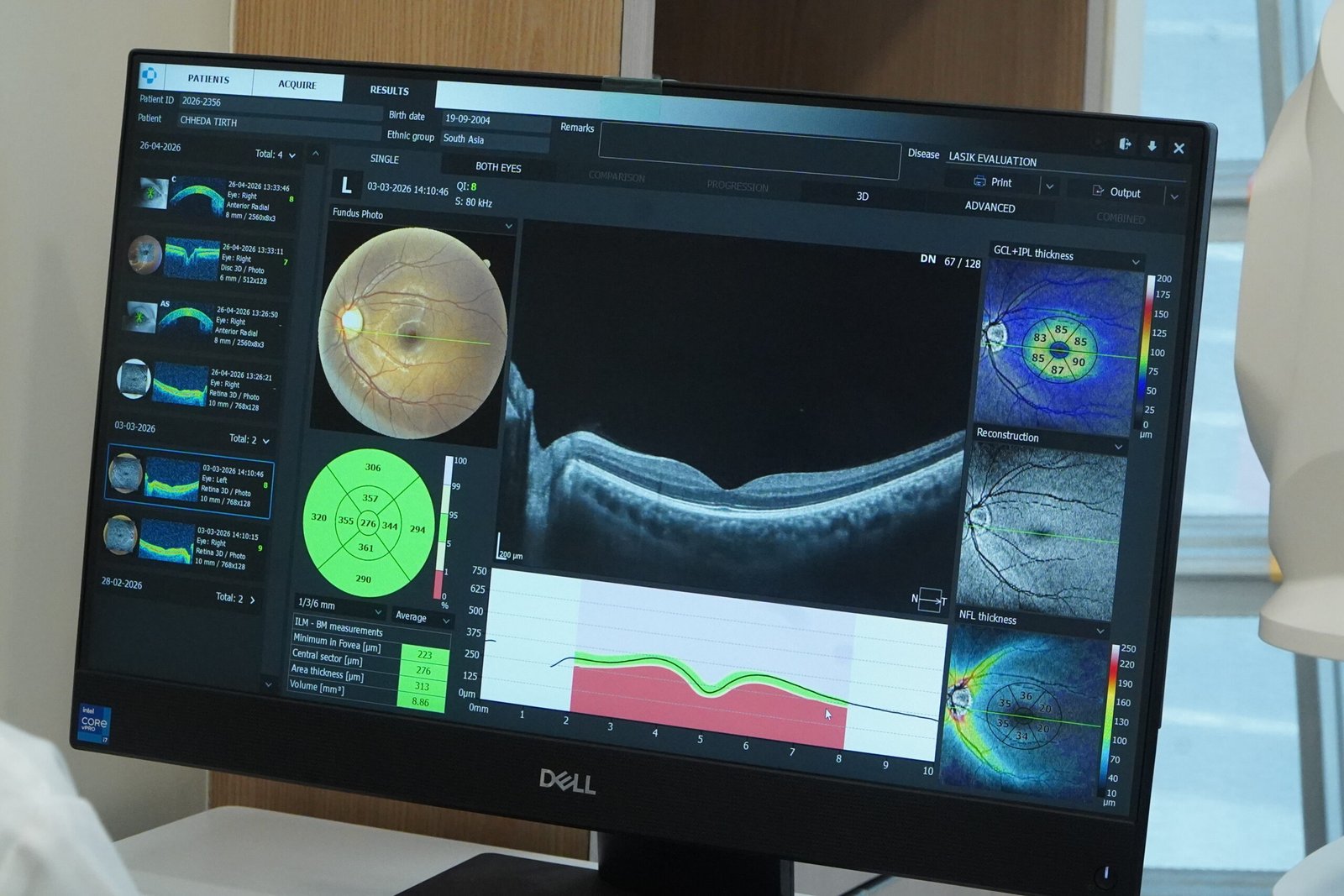
Here is the uncomfortable truth of Indian eye care: most cataract surgeons never train behind the lens, and most general ophthalmologists have never peeled a membrane. When a routine surgery turns complicated mid-table — the capsule ruptures, the nucleus sinks — the eye needs a posterior-segment surgeon in that room, in that minute. Dr. Gala’s dual fluency (cataract + vitreoretinal, FRCS Glasgow, L.V. Prasad fellowship) means:
- No mid-crisis referrals: the rescue happens in the same theatre, same admission, by the same surgeon who started — or by the surgeon other doctors call.
- Complete clearance: fragment removal, retinal check, and lens fixation finished together — not in three instalments across three clinics.
- Research-current technique: he is fully aware of the latest research unlike old practitioners — from 25-gauge platforms and Yamane fixation to the evolving endophthalmitis evidence (120+ references below, all 2018–2026).
5. Failed or Referred Cases — Second-Opinion Surgery Welcome
Had surgery elsewhere that didn’t work? Recurrent retinal detachment, persistent macular hole, dropped nucleus still waiting for stage two, an IOL that was never placed, vision worse after surgery than before — these are the cases Crystal Clear Eye Clinic and Topax Eye Hospital see every month, referred by patients and by other surgeons alike.
You will get: a complete re-evaluation (OCT + examination + review of prior records), an honest verdict on what is still fixable, a written surgical plan with realistic outcomes — and if surgery won’t help, you will be told that too, plainly. Surgeons reading this: referrals are respected, co-managed, and reported back. A complicated case is not a failure of the first surgeon — eyes are humbling — but leaving a patient without a plan is.
6. Honest Outcomes & Success Rates (2026)
Moorfields-style transparency — because you deserve numbers, not adjectives. These are evidence-supported ranges for experienced vitreoretinal hands; your individual estimate is given in writing after examination:
| Surgery | Anatomical Success (1 surgery) | Key Caveat |
|---|---|---|
| Retinal detachment (primary) | 90–95% | Macula-on cases keep far better vision; PVR lowers single-surgery success |
| Macular hole closure | 90–95% (85–90% large/chronic) | Vision improves over 3–6 months, rarely to 6/6 |
| ERM peeling | ~95% membrane removal | Distortion improves more than letter-chart vision |
| Diabetic tractional detachment | 85–90% | Visual ceiling set by pre-existing diabetic damage |
| Dropped nucleus rescue + secondary IOL | >90% completed in one admission | Delay and retained fragments worsen prognosis |
| Scleral-fixated IOL | >95% stable fixation | Requires complete vitrectomy first — non-negotiable |
| Endophthalmitis | Eye saved in majority when treated early | Hours matter; late presentation limits everything |
| Open-globe trauma | Varies widely by injury | Honest staging — some eyes are partial saves |
Any surgeon quoting 100% for complicated cases is quoting marketing, not medicine. What you are owed is a skilled surgeon, current technique, and honest numbers — that is the deal here.
7. Complicated Eye Surgery Cost in Mumbai (2026 Price Table)
Date-stamped (24 July 2026), Mumbai-wide ranges from published competitor pricing — and where Dr. Gala’s centres sit within them:
| Surgery | Mumbai Published Range | At Dr. Gala’s Centres |
|---|---|---|
| Vitrectomy (simple — hemorrhage/ERM) | ₹40,000–70,000 (aggregators quote ₹38,000–65,000 avg) | Within honest city ranges; written package estimate after evaluation — surgeon, anaesthetist, consumables and follow-ups itemized. Direct cashless via Topax Eye Care; TPA cashless via Crystal Clear Eye Clinic |
| Retinal detachment surgery | ₹50,000–1,50,000 (+silicone oil ₹60,000–1,20,000) | |
| Macular hole surgery | ₹45,000–1,20,000 | |
| Complex diabetic vitrectomy | ₹70,000–1,50,000 | |
| Scleral-fixated IOL / secondary IOL | ~₹70,000 + anaesthetist (the one published Mumbai benchmark) | Quoted individually — fixation method chosen per eye |
| Complex cataract surgery | No Mumbai competitor publishes a distinct price (standard cataract ₹25,000–95,000 by lens) | Transparent add-on pricing for hooks/rings/backup — no surprise “it became complicated” bills |
| Endophthalmitis / trauma | Emergency — quoted on presentation | Emergency-first policy: sight-saving never waits for paperwork |
Revision/re-surgery policy: where a re-operation is needed within the treatment plan, it is discussed and priced upfront — some Mumbai centres discount re-surgery up to 80%; ask for the policy in writing wherever you go, including here.
8. Insurance, Cashless & EMI
- Complicated surgeries are exactly what health insurance exists for — retinal detachment, vitrectomy, endophthalmitis and trauma repairs are covered procedures under most mediclaim policies (unlike OPD tests) — details on our cashless eye surgery page.
- Cashless split: direct cashless is done through Topax Eye Care (Vile Parle East); third-party/TPA cashless is done through Crystal Clear Eye Clinic (Andheri West). Bring your policy card; the team handles pre-authorization paperwork.
- CGHS / government schemes: documentation provided in claim-office formats.
- EMI options available for packages — ₹1,20,000 should never be the reason a retina stays detached. Ask on 077188 85245.
9. Why Dr. Jignesh Gala — Credentials & Difference
Patients searching for the best eye specialist in Mumbai, the best retina surgeon in Mumbai, or a complicated eye surgery specialist in Andheri West should check three things — training, volume, and honesty. Here is Dr. Gala’s card:
✅ Training: FRCS (Glasgow) • MRCS (Edinburgh) • FICO • Vitreoretinal fellowship at L.V. Prasad Eye Institute • founding ophthalmology team, Woodlands Health, Singapore • former Assistant Professor, B.Y.L. Nair Hospital — a municipal-hospital tenure where complicated cases are the norm, not the exception.
✅ He owns both centres — Crystal Clear Eye Clinic (Andheri West) and Topax Eye Hospital (Vile Parle East). No visiting-surgeon handoffs: the doctor who evaluates you operates you and follows you up — watch him work in his surgical videos.
✅ Peer-trusted: six years as a BMJ Case Reports peer reviewer — he evaluates other surgeons’ complex cases for an international journal.
✅ Fully aware of the latest research, unlike old practitioners — every technique on this page is backed by 2018–2026 PubMed evidence (library below): PIVOT-era detachment trials, inverted-flap macular hole data, modern endophthalmitis timing debates. He practises 2026’s surgery, not 2006’s.
✅ Front + back of the eye: complicated cases rarely respect anatomy — a surgeon who does both complex cataract and vitreoretinal work finishes the job in one admission.
10. Locations — Complicated Eye Surgery in Andheri West & Vile Parle East
1) Crystal Clear Eye Clinic — Andheri West
📍 Address: Laram Centre CHS, A1-202, Swami Vivekanand Road, above Sunil Jewellers, near NADCO Shopping Centre, Railway Colony, Andheri West, Mumbai 400058
📞 Phone: 077188 85245 • 🕘 Open daily, closes 9 pm
💳 Third-party / TPA cashless through Crystal Clear Eye Clinic.
Serving Andheri West, Lokhandwala, Versova, Yari Road, Juhu, Oshiwara, DN Nagar, Seven Bungalows, Azad Nagar, Veera Desai Road, Jogeshwari, Vile Parle West, Amboli — with complicated-surgery patients travelling in from Bandra, Khar, Santacruz, Goregaon, Malad, Kandivali, Borivali, Powai, Dadar, Thane and Navi Mumbai.
2) Topax Eye Hospital — Vile Parle East
📍 Vile Parle East, Mumbai (full address on drjigneshgalaeye.com) • 📞 077188 85245
💳 Direct cashless through Topax Eye Care. The hospital setting for major complicated surgeries — vitrectomy, trauma reconstruction, endophthalmitis — with admission, anaesthesia and post-op care under Dr. Gala’s own team. Convenient for Vile Parle, Santacruz East, Khar, Bandra East, Kalina, Vakola, Juhu and the airport corridor.

11. हिंदी • मराठी • ગુજરાતી — Patient Summaries
🇮🇳 हिंदी सारांश — जटिल आँख की सर्जरी
रेटिना अलग होना (रेटिनल डिटैचमेंट), मैक्युलर होल, डायबिटिक रेटिनोपैथी की सूजी नसें, सर्जरी के दौरान लेंस का अंदर गिरना, इंफेक्शन (एंडोफ्थैल्माइटिस) या चोट लगना — ये जटिल सर्जरियाँ किसी भी सामान्य सर्जन के बस की नहीं होतीं। डॉ. जिग्नेश गाला (FRCS Glasgow, L.V. प्रसाद फेलो) — अंधेरी वेस्ट, मुंबई में जटिल आँख की सर्जरी के विशेषज्ञ — खुद क्रिस्टल क्लियर आई क्लीनिक और टोपैक्स आई हॉस्पिटल में ये सर्जरियाँ करते हैं। कहीं और हुई नाकाम सर्जरी के बाद भी सलाह और री-सर्जरी उपलब्ध। लागत ₹40,000–1,50,000; कैशलेस सुविधा उपलब्ध। आपात स्थिति में तुरंत कॉल करें: 077188 85245।
🚩 मराठी सारांश — गुंतागुंतीची डोळ्यांची शस्त्रक्रिया
रेटिना सुटणे (रेटिनल डिटॅचमेंट), मॅक्युलर होल, मधुमेहामुळे डोळ्यात रक्तस्राव, शस्त्रक्रियेत लेन्स आत पडणे, डोळ्यातील इन्फेक्शन किंवा इजा — अशा गुंतागुंतीच्या शस्त्रक्रिया विशेष प्रशिक्षण मागतात. डॉ. जिग्नेश गाला (FRCS Glasgow, L.V. प्रसाद फेलो) — अंधेरी वेस्ट, मुंबई येथे गुंतागुंतीची डोळ्यांची शस्त्रक्रिया करणारे विट्रिओरेटिना सर्जन — स्वतःच्या क्रिस्टल क्लियर आय क्लीनिक व टोपॅक्स आय हॉस्पिटलमध्ये या शस्त्रक्रिया करतात. इतरत्र अयशस्वी शस्त्रक्रियेनंतरही दुसऱ्या मतासाठी व पुनःशस्त्रक्रियेसाठी येऊ शकता. खर्च ₹४०,०००–१,५०,०००; कॅशलेस सुविधा उपलब्ध. आपत्कालीन स्थितीत लगेच कॉल: 077188 85245.
🟡 ગુજરાતી સારાંશ — જટિલ આંખની સર્જરી
રેટિનલ ડિટેચમેન્ટ, મેક્યુલર હોલ, ડાયાબેટીસથી આંખમાં રક્તસ્રાવ, સર્જરી દરમિયાન લેન્સ અંદર પડવું, આંખનો ચેપ કે ઈજા — આવી જટિલ સર્જરીઓ ખાસ તાલીમ માગે છે. ડૉ. જિગ્નેશ ગાલા (FRCS Glasgow, L.V. પ્રસાદ ફેલો) — અંધેરી વેસ્ટ, મુંબઈમાં જટિલ આંખની સર્જરીના નિષ્ણાત — પોતાના ક્રિસ્ટલ ક્લિયર આય ક્લિનિક અને ટોપેક્સ આય હોસ્પિટલમાં આ સર્જરીઓ જાતે કરે છે. બીજે નિષ્ફળ સર્જરી થઈ હોય તો પણ બીજા મત અને ફરી સર્જરી માટે આવી શકો છો. ખર્ચ ₹૪૦,૦૦૦–૧,૫૦,૦૦૦; કેશલેસ સુવિધા ઉપલબ્ધ. ઈમર્જન્સીમાં તરત કૉલ: 077188 85245.
12. ❓ 12 Frequently Asked Questions (English + Hinglish)
1. Complicated eye surgery mein risk kitna hota hai? (How risky is it?)
More than routine surgery, less than fear suggests. In experienced vitreoretinal hands: primary retinal detachment repair succeeds in 90–95% with one operation; macular holes close in 90–95%; serious infection risk is under 1 in 1,000. The real risk is delay — complicated eyes worsen waiting. You will always get your personal risk estimate in writing before consent.
2. Retinal detachment surgery ke baad vision kitna wapas aata hai?
Depends on one thing more than any other: was your macula still attached when you arrived? Macula-on detachments often recover near-full vision; macula-off cases recover partially over 3–6 months. That is why a curtain/shadow in vision is a same-day emergency — call 077188 85245 immediately.
3. What is the cost of complicated eye surgery in Mumbai?
Published 2026 Mumbai ranges: simple vitrectomy ₹40,000–70,000; retinal detachment ₹50,000–1,50,000; macular hole ₹45,000–1,20,000; complex diabetic vitrectomy ₹70,000–1,50,000; scleral-fixated IOL ~₹70,000. At our centres you get an itemized written package before surgery (see surgery packages) — no surprise bills. Cashless and EMI available.
4. Meri pehli surgery fail ho gayi – kya dobara surgery ho sakti hai?
Ha, aksar ho sakti hai. Recurrent retinal detachment, persistent macular hole, dropped nucleus ke remnants, dislocated IOL — revision surgery is a large part of our complex-case work. Pehle complete re-evaluation (OCT + records review), phir honest verdict: kya fixable hai, kitna vision realistic hai, aur kya plan hoga. Failed surgery ka patient = hamara priority patient.
5. Macular hole surgery ke baad face-down position kyun rakhna padta hai?
Gas bubble ko macula par press rehna hota hai taaki hole band ho — isliye kuch din face-down (ya specific position) advise hoti hai. Modern evidence shows positioning requirements are shorter and gentler than old protocols — and some holes need none. Aapko aapke hole ke hisaab se exact instructions milenge, likhit form mein.
6. How long is recovery after a vitrectomy?
Eye feels scratchy 1–2 weeks; vision is blurry while gas is inside (2–8 weeks depending on gas type; silicone oil stays longer and is removed later). Desk work usually resumes in 2–3 weeks; flying is forbidden while gas is in the eye. Final vision settles over 3–6 months. Follow-ups: day 1, week 1, then monthly initially.
7. Dropped nucleus ke baad kitne din mein surgery karani chahiye?
Ideally within days, not weeks. Retained lens material causes inflammation, pressure spikes and retinal damage the longer it stays. If your cataract surgery elsewhere ended with “the lens has fallen back,” ask for a vitreoretinal referral the same week — or call us directly. Complete removal + secondary IOL is usually one admission.
8. Is complicated eye surgery covered by insurance / cashless?
Yes — retinal detachment, vitrectomy, endophthalmitis and trauma surgeries are covered under most mediclaim policies (unlike OPD tests). Direct cashless via Topax Eye Care; TPA/third-party cashless via Crystal Clear Eye Clinic. Our team handles pre-authorization. EMI options exist for uncovered portions.
9. Kya diabetes wale patient ki retina surgery safe hai?
Safe aur zaroori — lekin planning maangti hai. Sugar control, kidney status aur blood thinners pehle optimize hote hain; anti-VEGF injection se surgery aasaan ho jaati hai. Diabetic tractional detachment mein anatomical success 85–90% tak hai modern techniques se. Surgery ke baad sugar control = aadha ilaaj, ye samajhna zaroori hai.
10. Complicated eye surgery kahan karwaye Andheri West mein?
Crystal Clear Eye Clinic — Laram Centre CHS, A1-202, S.V. Road, above Sunil Jewellers, near NADCO Shopping Centre, Andheri West 400058 (evaluation + lasers); Topax Eye Hospital, Vile Parle East (major surgeries + admission). Same phone: 077188 85245. Open daily, closes 9 pm. Emergencies seen same day.
11. Silicone oil kya hota hai aur kab nikala jata hai?
Complicated detachments mein retina ko andar se support dene ke liye silicone oil bhara jata hai — jaise internal splint. Ye temporary hai: usually 2–6 months mein ek chhoti second surgery se nikala jata hai (Mumbai benchmark: removal ~₹25,000). Oil ke andar vision blurry rehti hai; removal ke baad improve hoti hai. Gas ke comparison mein oil lambi support deta hai — isliye complicated cases mein use hota hai.
12. How do I know if my case needs a retina specialist or a general eye doctor?
Simple test: anything behind the lens (retina, vitreous, macula, detachment, diabetic bleeding, dropped lens material, eye infections inside) = vitreoretinal territory. Anything routine in front (simple cataract, glasses, surface problems) = general. And the grey zone — complicated cataracts, dislocated lenses, failed surgeries — goes to the surgeon who does both. A 5-minute call to 077188 85245 with your reports gets you the right answer.
13. 📖 Related Reading — Complete Eye-Care Library by Dr. Jignesh Gala (78 guides)
Every guide below is written by the same surgeon who would operate on you:
From drjigneshgalaeye.com
- Drjigneshgalaeye.com
- A year at l v prasad eye institute how my vitreoretinal fellowship shaped the surgeon i am today
- About us
- Anti vegf injection cost in mumbai 2026 every drug every price explained honestly by a retina specialist
- B y l nair hospital 2019 2020 my sixteen months as assistant professor of ophthalmology
- Bakuchiol eye cream a gentle revolution in under eye care
- Best eye doctor for retina in mumbai dr jignesh gala retina vitreoretinal surgeon
- Best eye specialist in andheri mumbai
- Best retina specialist mumbai dr jignesh gala
- Best vision correction surgery after 40 what are your options
- Blogs by dr jignesh gala
- Cashless eye surgery in mumbai dr jignesh gala
- Diabetic retinopathy specialist in andheri west mumbai advanced screening injections laser vitrectomy by dr jignesh gala frcs
- Discover crystal clear vision with contoura lasik in mumbai
- Dr jignesh gala top eye specialist in mumbai
- Drjignesh gala premier pediatric ophthalmologistandheri mumbai
- Eylea injection in mumbai cost uses side effects the 2026 price revolution explained by a retina specialist
- Glaucoma treatment in andheri mumbai
- How to prevent dry eyes in winter
- Hvf test in andheri west mumbai visual field test cost procedure how to read your report by a specialist
- I see a curtain or shadow in my vision
- Inside my six years as a bmj case reports peer reviewer
- Inside the fico 2016 the only worldwide medical specialty examination
- Inside the frcs ophth glasgow how a 425 year old royal college tests your surgical knowledge
- Intravitreal injection in andheri west mumbai painless anti vegf eye injections by dr jignesh gala frcs
- Know your eye doctor
- Laser cataract surgery in andheri mumbai dr jignesh gala
- Laser cataract surgery
- Lasik eye surgery in andheri mumbai dr jignesh gala
- Lasik eye surgery in bandra mumbai dr jignesh gala
- Lasik eye surgery in juhu dr jignesh gala
- Lasik eye surgery in lokhandwala dr jignesh gala
- Lasik eye surgery in oshiwara dr jignesh gala
- Lasik in santacruz mumbai
- Lasik in versova mumbai dr jignesh gala
- Lasik surgery in andheri mumbai
- Most common types of refractive eye surgery which one is right for you
- Mrcs edinburgh how i joined a royal college founded in 1505
- Multifocal trifocal iol guide complete patient guide
- Oct eye test in andheri west mumbai cost procedure report why the specialist reading it matters
- People do ask google about this eye issue
- Refraction in real time how the royal college of ophthalmologists tested me in kuching 2017
- Refractive lens exchange rlethe complete guide to clear vision without glasses
- Retina laser treatment in andheri west mumbai prp barrage focal micropulse laser by dr jignesh gala frcs
- Retina surgery in mumbai your complete guide to saving your sight
- Retina treatment in andheri
- Retina treatment in mumbai advanced retinal care by dr jignesh gala
- Rle vs lasik which vision correction surgery is better
- Satisfied patient stories
- Singapore 2021 a year on the founding ophthalmology team of woodlands health
- Smartsurf with customeyes the most advanced glass removal surgery in mumbai and why it outperforms wavelight plus innoveyes
- Surgery packages
- Surgical videos by dr jignesh gala
- Types of intraocular lenses
- Types of refractive surgeries
- Types of retina surgeries
- Understanding and preventing digital eye strain
From crystalcleareye.in
- Crystal Clear Eye Clinic — Home
- About Crystal Clear Eye Clinic, Andheri West
- Eye Health Blog — Crystal Clear Eye Clinic
- Contact & Directions — Andheri West
- Glaucoma Treatment in Andheri, Mumbai
- LASIK Surgery in Andheri, Mumbai
- Premium IOL Selection Guide 2026
- Laser Cataract Surgery in Andheri, Mumbai
- Best Eye Specialist in Andheri, Mumbai
- Best Retina Specialist in Mumbai
- Best Vitrectomy Surgery in Mumbai — Expert Retinal Care
- Diabetic Eye Care in Mumbai
- Diabetic Retinopathy in Andheri
- Diabetic Macular Edema (DME) Treatment in Mumbai
- Macular Edema Treatment in Andheri West
- ARMD Treatment in Andheri West, Mumbai
- Retina Treatment in Andheri
- Vitreoretinal Treatment in Andheri
- Cashless Eye Surgery in Mumbai
- Sudden Floaters? Retina Check-up in Mumbai
- What Is an Intravitreal Eye Injection?
Complicated case? This is exactly the clinic you were looking for.
Whether you searched for a complicated eye surgery in Mumbai, the best eye specialist in Mumbai for a difficult case, a complex cataract surgeon in Andheri West, or a second opinion after failed eye surgery — you have found a surgeon who trains internationally, publishes-grade reads the evidence, owns his hospitals, and answers his own phone line.
📍 Andheri West: Laram Centre CHS, A1-202, S.V. Road, above Sunil Jewellers, near NADCO Shopping Centre, Mumbai 400058
📍 Vile Parle East: Topax Eye Hospital • Open daily, closes 9 pm • Retinal emergencies same-day
Dr. Jignesh Gala, FRCS (Glasgow) — Vitreoretinal & Cataract Surgeon • drjigneshgalaeye.com • crystalcleareye.in
Medical disclaimer: This page is educational and does not replace a personal consultation. Costs are indicative Mumbai ranges verified on 24 July 2026; your written estimate follows evaluation. Outcome figures are population-level evidence, not guarantees — every complicated eye is its own story, and yours will be told to you honestly.
14. Research & Evidence Library — 120+ Verified References (2018–2026)
Every technique on this page is backed by recent, verified PubMed research (all 2018–2026, harvested via official NCBI E-utilities in July 2026). This is the evidence Dr. Gala reads — click any title to verify the science yourself.
Pars Plana Vitrectomy For Rhegmatogenous Retinal Detachment
- Retinal detachment. — Nature reviews. Disease primers, 2024
- The Pneumatic Retinopexy versus Vitrectomy for the Management of Primary Rhegmatogenous Retinal Detachment Outcomes Randomized Trial (PIVOT). — Ophthalmology, 2019
- [Scleral Buckling]. — Klinische Monatsblatter fur Augenheilkunde, 2019
- Early Versus Delayed Vitrectomy for Vitreous Hemorrhage Secondary to Proliferative Diabetic Retinopathy. — American journal of ophthalmology, 2025
- Outcomes Of Microincision Pars Plana Vitrectomy In Rhegmatogenous Retinal Detachment. — Journal of Nepal Health Research Council, 2023
- Pars Plana Vitrectomy Outcomes for Rhegmatogenous Retinal Detachment Qualifying for Pneumatic Retinopexy. — Clinical ophthalmology (Auckland, N.Z.), 2021
- PARS PLANA VITRECTOMY-SUPRACHOROIDAL VISCOPEXY FOR RHEGMATOGENOUS RETINAL DETACHMENT REPAIR. — Retina (Philadelphia, Pa.), 2026
- Pars Plana Vitrectomy in the Treatment of Rhegmatogenous Retinal Detachment. — Ceska a slovenska oftalmologie : casopis Ceske oftalmologicke spolecnosti a Slovenske oftalmologicke spolecnosti, 2024
- Retinal Displacement after Pneumatic Retinopexy versus Vitrectomy for Rhegmatogenous Retinal Detachment (ALIGN). — Ophthalmology, 2022
- Cystoid macular oedema following pars plana vitrectomy for macula on rhegmatogenous retinal detachment. — Clinical & experimental optometry, 2021
- Pneumatic retinopexy: an update. — Graefe’s archive for clinical and experimental ophthalmology = Albrecht von Graefes Archiv fur klinische und experimentelle Ophthalmologie, 2022
- Rhegmatogenous Retinal Detachment in Childhood. — Ceska a slovenska oftalmologie : casopis Ceske oftalmologicke spolecnosti a Slovenske oftalmologicke spolecnosti, 2025
- Comparison of Scleral Buckling and Vitrectomy Using Wide Angle Viewing System for Rhegmatogenous Retinal Detachment. — Seminars in ophthalmology, 2020
- Outcomes of Pars Plana Vitrectomy in Inherited Retinal Degenerations. — Ophthalmology. Retina, 2026
- Pneumatic Retinopexy Versus Pars Plana Vitrectomy for Rhegmatogenous Retinal Detachment: Systematic Review and Meta-Analysis. — Ophthalmic surgery, lasers & imaging retina, 2023
- Rhegmatogenous Retinal Detachment after Intravitreal Injection. — Ophthalmology. Retina, 2021
- Pneumatic retinopexy: A critical reappraisal. — Survey of ophthalmology, 2021
- Safety and efficacy of cryopexy during pars Plana vitrectomy in rhegmatogenous retinal detachment. — European journal of ophthalmology, 2023
- Combined pars plana vitrectomy with phacoemulsification for rhegmatogenous retinal detachment repair. — Clinical ophthalmology (Auckland, N.Z.), 2019
- RETINAL DISPLACEMENT AFTER RHEGMATOGENOUS RETINAL DETACHMENT REPAIR: Scleral Buckling versus Pars Plana Vitrectomy (The BEVERLEY Study). — Retina (Philadelphia, Pa.), 2025
Macular Hole Surgery: Inverted Flap & Ilm Peeling
- Complete ILM Peeling Versus Inverted Flap Technique for Macular Hole Surgery: A Meta-Analysis. — Ophthalmic surgery, lasers & imaging retina, 2020
- Innovative Non-Inverted ILM Free Flap Covering Technique for Unclosed Macular Hole Repair. — American journal of ophthalmology, 2026
- Internal limiting membrane peeling in macular hole surgery. — German medical science : GMS e-journal, 2022
- Refractory Macular Hole Surgery: A Review of Recent Surgical Innovations. — International ophthalmology clinics, 2025
- Anatomical prognosis after idiopathic macular hole surgery: machine learning based-predection. — The Libyan journal of medicine, 2022
- [Large macular hole-Always a poor prognosis?]. — Der Ophthalmologe : Zeitschrift der Deutschen Ophthalmologischen Gesellschaft, 2021
- Modified inverted internal limiting membrane flap technique for macular hole closure. — Japanese journal of ophthalmology, 2022
- Comparison of Techniques: Inverted Flap and Conventional Internal Limiting Membrane Removal in Idiopathic Macular Hole Surgery. — Ceska a slovenska oftalmologie : casopis Ceske oftalmologicke spolecnosti a Slovenske oftalmologicke spolecnosti, 2024
- Inverted Internal Limiting Membrane Flap versus Internal Limiting Membrane Peeling for <400 μm Macular Hole: A Meta-Analysis and Systematic Review. — Ophthalmic research, 2023
- Anatomical and Functional Outcomes in Eyes with Idiopathic Macular Holes that Underwent Surgery Using the Inverted Internal Limiting Membrane (ILM) Flap Technique Versus the Conventional ILM Peeling Technique. — Advances in therapy, 2021
- Outcomes of Vitrectomy for Long-Duration Macular Hole. — Journal of clinical medicine, 2020
- Inverted ILM Flap Technique in Idiopathic Full-Thickness Macular Hole Surgery: Functional Outcomes and Their Correlation with Morphologic Findings. — Journal of ophthalmology, 2021
- Modification of the inverted flap technique compared to conventional internal limiting membrane peeling in complete macular hole surgery. — European journal of ophthalmology, 2023
- Impact of Epiretinal Membrane on Anatomical and Visual Outcomes in Patients with Full-Thickness Macular Holes. — Ophthalmology. Retina, 2025
- Anatomical and functional results after vitrectomy with conventional ILM peeling versus inverted ILM flap technique in large full-thickness macular holes. — International journal of retina and vitreous, 2023
- Dissociated Optic Nerve Fiber Layer Appearance after Macular Hole Surgery: A Randomized Controlled Trial Comparing the Temporal Inverted Internal Limiting Membrane Flap Technique with Conventional Peeling. — Ophthalmology. Retina, 2023
- Vitrectomy with inverted internal limiting membrane flap versus internal limiting membrane peeling for macular hole retinal detachment in high myopia: a systematic review of literature and meta-analysis. — Eye (London, England), 2019
- Internal Limiting Membrane Flap and Insertion Techniques Improve Prognosis in Macular Hole-Associated Retinal Detachment. — Ophthalmology. Retina, 2026
- Myopic traction maculopathy in fovea-involved myopic chorioretinal atrophy. — Eye (London, England), 2024
- Best surgical technique and outcomes for large macular holes: retrospective multicentre study in Japan. — Acta ophthalmologica, 2018
Diabetic Vitrectomy: Tractional Retinal Detachment & Vitreous Hemorrhage
- Initial Therapy of Panretinal Photocoagulation vs Anti-VEGF Injection for Proliferative Diabetic Retinopathy. — JAMA ophthalmology, 2024
- Tractional Retinal Detachment Secondary to Diabetic Retinopathy. — JAMA ophthalmology, 2018
- Pharmacological adjuvants for diabetic vitrectomy surgery. — World journal of methodology, 2024
- Vitrectomy for cases of diabetic retinopathy. — Indian journal of ophthalmology, 2024
- Post-vitrectomy delayed retinal breaks in proliferative diabetic retinopathy. — International journal of retina and vitreous, 2023
- Tractional Retinal Detachment in Eyes with Vitreous Hemorrhage and Proliferative Diabetic Retinopathy and Posterior Vitreous Detachment in Fellow Eye. — Korean journal of ophthalmology : KJO, 2023
- Analysis of Risk Factors for Revitrectomy in Eyes with Diabetic Vitreous Hemorrhage. — Diabetes, metabolic syndrome and obesity : targets and therapy, 2023
- Outcomes and Complications of Pars Plana Vitrectomy for Nonclearing Vitreous Hemorrhage Without Tractional Elements. — Ophthalmology. Retina, 2026
- Vitrectomy for diabetic retinopathy: A review of indications, techniques, outcomes, and complications. — Taiwan journal of ophthalmology, 2024
- Individual and Systems-Based Risk Factors for Diabetic Vitrectomy in an Urban Safety-Net Hospital. — Ophthalmology. Retina, 2023
- Postoperative Complications of Pars Plana Vitrectomy for Diabetic Retinal Disease. — Seminars in ophthalmology, 2018
- Elucidating postoperative dynamics in tractional retinal detachment: a systematic review and meta-analysis of structural and functional outcomes following diabetic vitrectomy, including an analysis of postoperative complications. — BMC ophthalmology, 2024
- Preoperative Evaluation of Tractional Retinal Detachment with B-Mode Ultrasonography in Diabetic Vitreous Hemorrhage. — Beyoglu eye journal, 2021
- The Anatomical and Functional Outcomes of 27-Gauge Pars Plana Vitrectomy in Diabetic Tractional Retinal Detachments in the South Asian Population. — Cureus, 2023
- Tips, Tricks, and Advantages of 27-G Vitrectomy. — Ophthalmologica. Journal international d’ophtalmologie. International journal of ophthalmology. Zeitschrift fur Augenheilkunde, 2018
- Commentary: Outcomes of preoperative bevacizumab in diabetics with nonclearing vitreous hemorrhage without tractional detachment – A quasi-randomized retrospective study. — Indian journal of ophthalmology, 2021
- [Medico-surgical management of intravitreal hemorrhage in diabetic patients]. — Journal francais d’ophtalmologie, 2023
- ALL-PROBE VITRECTOMY DISSECTION TECHNIQUES FOR DIABETIC TRACTIONAL RETINAL DETACHMENTS: Lift and Shave. — Retina (Philadelphia, Pa.), 2018
- SURGICAL OUTCOMES IN CONCURRENT SICKLE CELL AND DIABETIC RETINOPATHY. — Retina (Philadelphia, Pa.), 2024
- Impact of COVID-19 lockdown on Retinal Surgeries. — Pakistan journal of medical sciences, 2021
Complex Cataract Surgery: Subluxated Lens, Posterior Polar, Small Pupil
- Pseudoexfoliation Glaucoma: Clinical Presentation and Therapeutic Options. — Turkish journal of ophthalmology, 2023
- Iridoschisis-A Systematic Review. — Journal of clinical medicine, 2020
- Descemet membrane detachment. — Survey of ophthalmology, 2020
- Slow swirl phaco. — Indian journal of ophthalmology, 2024
- The escape route: novel surgical technique for the posterior polar cataract. — Journal of cataract and refractive surgery, 2022
- Future Intraocular Lens Technologies. — Ophthalmology, 2021
- Causes of intraocular pseudophakic lens explantation in Spain. — European journal of ophthalmology, 2024
- Late in-the-bag intraocular lens dislocation. — Journal of cataract and refractive surgery, 2021
- Dilation devices in cataract surgery. — Current opinion in ophthalmology, 2023
- “Raw egg”: Hypermature cataract with lens dislocation. — Journal francais d’ophtalmologie, 2023
- Cataract surgery in retinitis pigmentosa. — Medical hypothesis, discovery & innovation ophthalmology journal, 2024
- Phacoemulsification of posterior polar cataracts. — Journal of cataract and refractive surgery, 2019
- Diagnosis and treatment of microspherophakia. — Journal of cataract and refractive surgery, 2020
- Management of the subluxated crystalline lens: A review. — Clinical & experimental ophthalmology, 2021
- Pharmacological management of intra-operative miosis during cataract surgery. — Indian journal of ophthalmology, 2023
- Indications for surgical management of retained lens fragments. — Current opinion in ophthalmology, 2022
- Intraocular lens exchange or explantation post cataract surgery. — International journal of ophthalmology, 2024
- Intracameral medications in cataract surgery. — International ophthalmology, 2025
- Crystalline lens alterations in congenital aniridia. — Archivos de la Sociedad Espanola de Oftalmologia, 2021
- Approach to a spontaneously ruptured posterior polar cataract. — Journal of cataract and refractive surgery, 2023
Scleral-Fixated Iol & Dropped Nucleus Management
- Intraocular lens implantation in the absence of capsular support: scleral fixation. — Eye (London, England), 2022
- My X-nit. — Indian journal of ophthalmology, 2023
- Artificial iris exchange. — Journal of cataract and refractive surgery, 2020
- Scleral fixation of intraocular lenses. — Current opinion in ophthalmology, 2020
- Complications of Scleral-Fixated Intraocular Lenses. — Seminars in ophthalmology, 2018
- Scleral-fixated and iris-fixated intraocular lens implantation or fixation:meta-analysis. — Journal of cataract and refractive surgery, 2022
- Loose zonules in cataract surgery. — Current opinion in ophthalmology, 2022
- Frugal model for scleral fixated intraocular lens simulation. — Indian journal of ophthalmology, 2021
- Complications of Secondary Scleral-fixated Intraocular Lens Placement. — International ophthalmology clinics, 2022
- Aetiology and Management of IOL Dislocations. — Klinische Monatsblatter fur Augenheilkunde, 2023
- Haptic Erosion Following Sutureless Scleral-fixated Intraocular Lens Placement. — Ophthalmology. Retina, 2023
- Retropupillary Iris-fixated versus Sutured Scleral-fixated Intraocular Lenses. — Romanian journal of ophthalmology, 2024
- Refractive Outcomes of the Yamane Flanged Intrascleral Haptic Fixation Technique. — Ophthalmology, 2020
- Challenges with foldable intraocular lenses with hollow haptics or eyelets in scleral fixation. — Journal of cataract and refractive surgery, 2021
- Double-flanged polypropylene technique: 5-year results. — Journal of cataract and refractive surgery, 2023
- Outcomes and complications in scleral-fixated intraocular lens implantations. — International ophthalmology, 2020
- [Intraoperative opacification of a scleral-fixated Carlevale (Soleko®) intraocular lens]. — Journal francais d’ophtalmologie, 2024
- Scleral-fixated intraocular lens implants-evolution of surgical techniques and future developments. — Eye (London, England), 2021
- Flanged fixation: Yamane technique and its application. — Current opinion in ophthalmology, 2021
- Scleral fixated secondary intraocular lenses: a review of recent literature. — Current opinion in ophthalmology, 2020
Endophthalmitis Vitrectomy & Open Globe Repair
- When is vitrectomy for endophthalmitis needed? — Acta ophthalmologica, 2023
- Early Vitrectomy for Endophthalmitis: Are EVS Guidelines Still Valid? — Ophthalmic research, 2023
- Endophthalmitis. — British journal of hospital medicine (London, England : 2005), 2019
- Exogenous Endophthalmitis. — Ocular immunology and inflammation, 2023
- Endogenous Endophthalmitis. — The New England journal of medicine, 2023
- Endophthalmitis Prevention. — Asia-Pacific journal of ophthalmology (Philadelphia, Pa.), 2018
- [Endogenous Candida endophthalmitis]. — Die Ophthalmologie, 2024
- Correspondence. — Retina (Philadelphia, Pa.), 2019
- [Endophthalmitis]. — Der Ophthalmologe : Zeitschrift der Deutschen Ophthalmologischen Gesellschaft, 2018
- Early vitrectomy for severe eye injuries. — Eye (London, England), 2021
- Complete and early vitrectomy for endophthalmitis. — European journal of ophthalmology, 2021
- First endogenous fungal endophthalmitis due to Fusarium dimerum: A severe eye infection contracted during induction chemotherapy for acute leukemia. — Journal de mycologie medicale, 2018
- Endophthalmitis: Then and Now. — American journal of ophthalmology, 2018
- Reply. — Retina (Philadelphia, Pa.), 2019
- Vitreoretinal Surgery in Glaucoma. — Klinische Monatsblatter fur Augenheilkunde, 2022
- Pediatric Pars Planitis: A Review. — Ocular immunology and inflammation, 2023
- Complications of vitreoretinal surgery. — Current opinion in ophthalmology, 2020
- Ultrasound-Guided Pars Plana Vitrectomy. — Retina (Philadelphia, Pa.), 2023
- Endophthalmitis Associated With XEN Stent Implantation. — American journal of ophthalmology, 2023
- Postpartum Endogenous Candida Endophthalmitis. — Middle East African journal of ophthalmology, 2019