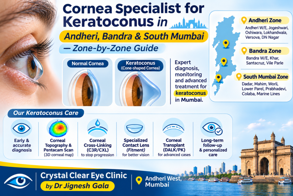
Cornea Specialist for Keratoconus in Andheri, Bandra & South Mumbai — Zone-by-Zone Guide
Written & medically reviewed by Dr. Jignesh Gala, Vitreoretinal & Cataract Surgeon (FRCS Glasgow), Founder & Director — Crystal Clear Eye Clinic (Andheri West) and Topax Eye Hospital (Vile Parle East), Mumbai. Facts verified: 24 July 2026.
Thank you for reading this post, don't forget to subscribe!⚡ Quick answer: Searching for a cornea specialist for keratoconus in Andheri or Bandra? Keratoconus is a follow-up disease — year one alone means 6–12 visits — so the best specialist is usually the fully equipped one nearest to you, not the most famous one across the city. For the western suburbs, Dr. Jignesh Gala (FRCS Glasgow) at Crystal Clear Eye Clinic, Andheri West offers corneal tomography on-site, serial progression records, cross-linking (C3R/CXL) at the centre, scleral-lens fitting with specialist fitters, and Intacs planning — with Topax Eye Hospital, Vile Parle East as the hospital arm. Call 077188 85245.
- Discover crystal clear vision with contoura lasik in mumbai
- Dr jignesh gala top eye specialist in mumbai
- Drjignesh gala premier pediatric ophthalmologistandheri mumbai
- Eylea injection in mumbai cost uses side effects the 2026 price revolution explained by a retina specialist
- Glaucoma treatment in andheri mumbai
- How to prevent dry eyes in winter
- Hvf test in andheri west mumbai visual field test cost procedure how to read your report by a specialist
- I see a curtain or shadow in my vision
- Inside my six years as a bmj case reports peer reviewer
- Inside the fico 2016 the only worldwide medical specialty examination
- Inside the frcs ophth glasgow how a 425 year old royal college tests your surgical knowledge
- Intravitreal injection in andheri west mumbai painless anti vegf eye injections by dr jignesh gala frcs
1. Zone-by-Zone: Andheri, Bandra, South Mumbai
Mumbai’s eye care is zonal, whether hospitals admit it or not. Here is the honest map for a keratoconus patient deciding where to anchor care:
| Zone | What You Find | Best For |
|---|---|---|
| Andheri West / East | Full keratoconus stack at Dr. Gala’s centres: tomography, progression records, CXL, scleral-lens coordination, Intacs planning. Minutes from Lokhandwala, Versova, Juhu, Oshiwara, DN Nagar, Jogeshwari. | Western-suburbs patients (Andheri to Kandivali) who need repeated scans and lens visits |
| Bandra / Khar / Santacruz | Good general ophthalmology; fewer centres with tomography + in-house CXL. Andheri West is 20–35 minutes by road or one train stop or two. | Bandra patients who want complete keratoconus care without a South Mumbai trek |
| South Mumbai | Big-name institutions and transplant-level corneal surgeons. | Genuine transplant-level disease and rare complications — worth the trip then, not for every scan |
| Vile Parle / airport corridor | Topax Eye Hospital (Vile Parle East) — Dr. Gala’s hospital arm, direct cashless. | Vile Parle, Santacruz East, Kalina, Khar East patients |
The pattern we see weekly: a Bandra or Juhu patient gets diagnosed at a famous South Mumbai institution, then quietly stops attending follow-ups because each visit costs half a day. Keratoconus punishes exactly that — progression is only provable from serial tomography (Kmax and ABCD grading compared over time), and missed scans mean missed progression windows. Diagnosis and treatment advances now favour early, monitored intervention (2023 review of keratoconus diagnosis & treatment).
2. The Follow-Up Commute Math
Do the arithmetic before choosing your specialist:
- Year one visits: diagnosis + baseline tomography, progression check at 3–6 months, CXL (if progressing) with 1-week, 1-month and 3–6-month reviews, plus 2–4 scleral-lens fitting visits. Realistic total: 6–12 visits.
- Time cost per visit: a specialist 15 minutes away = under 2 hours door-to-door. A ‘famous’ specialist 90 minutes away = 4–5 hours, half-day leave, and a strong temptation to skip.
- The clinical cost of skipping: pediatric and young-adult keratoconus progresses fastest — the exact patients whose parents postpone follow-ups. CXL works best when timed to documented progression, not to a convenient year later.
- Records continuity: scans must be compared on the same machine. Five visits at one Andheri West centre beat five visits at five ‘top’ hospitals whose machines cannot talk to each other.
The honest rule: for monitoring, lenses and CXL, the best cornea specialist is the well-equipped one you will actually visit. For a one-time transplant decision, travel is cheap. Keratoconus care is 90% the former.
3. The Andheri West Hub: What Dr. Gala’s Clinic Offers Keratoconus Patients
Everything keratoconus needs between diagnosis and the transplant question, in one Andheri West location:
- Corneal tomography/topography on-site — ₹1,200–2,500 per scan is the Mumbai band (advanced tomography up to ₹5,000; verified 24 July 2026). RGP wearers: 2-week lens washout before scanning for accurate readings.
- Progression records that trend — every scan stored and compared against your baseline with Kmax/ABCD grading. You see the numbers, not just an opinion.
- Cross-linking (C3R/CXL) at the centre — ₹25,000–45,000 per eye typical (published Mumbai range ₹20,000–90,000). CXL halts progression; it does not reverse the cone — so timing matters.
- Scleral-lens fitting coordinated with specialist fitters — scleral lenses vault the cornea entirely, and modern mini-sclerals rehabilitate even scarred and post-surgical corneas (BMC Ophthalmology 2025, scarred-cornea mini-sclerals 2024). Mumbai band: ₹15,000–50,000 per pair + fitting ₹500–3,000.
- Intacs evaluation & planning — ring segments improve lens tolerance in selected cases (₹60,000–1,20,000 per eye); they do not stop progression, so we plan them with CXL, not instead of it.
- Honest escalation — only ~10–15% of keratoconus ever needs a transplant. When that’s truly you, we say so plainly and co-manage the referral.
Full-service context matters too: keratoconus patients later need cataract and retinal care, and Dr. Gala’s centres cover the spectrum — from laser cataract surgery in Andheri to the subspecialty pages on types of retina surgeries. Our eye health blog documents the same evidence-first approach across conditions.
4. When Travelling IS Worth It
We are not saying “never cross the city.” Travel earns its hours when:
- Transplant-level disease: extreme thinning, deep scarring after hydrops, or corneas no lens can vault. DALK/PK is a one-time, surgeon-dependent decision — worth Mumbai’s best transplant hands, wherever they sit.
- Rare complications: unresolved hydrops, infectious ulcers in a keratoconic cornea, failed grafts.
- True second opinions on irreversible advice — before any big surgery, a fresh tomography-based review is worth any drive.
And here is how we make that work: for genuine transplant cases, Dr. Gala prepares the complete record set (serial tomography, refraction history, lens trials), refers to a corneal transplant specialist, and takes you back for the long follow-up — suture checks, surface care, and the scleral-lens rehabilitation that often follows grafts (mini-sclerals after penetrating keratoplasty, SPOT scleral lenses in keratoconus). Visual rehabilitation after any corneal surgery is a planned phase, not an afterthought (refractive rehabilitation review). You get the specialist surgery and the 15-minute follow-up.
5. Why Western-Suburbs Patients Choose Dr. Jignesh Gala

Dr. Jignesh Gala, FRCS (Glasgow), MRCS (Edinburgh), FICO — Founder & Director of Crystal Clear Eye Clinic (Andheri West) and Topax Eye Hospital (Vile Parle East); LVPEI fellowship; Woodlands Health Singapore founding team; ex-Assistant Professor, B.Y.L. Nair Hospital; 6 years a BMJ Case Reports peer reviewer. Learn more on the about page of Crystal Clear Eye Clinic or his profile as a leading eye specialist in Andheri.
✅ Location math on your side: two centres covering Andheri West to Vile Parle East — Lokhandwala, Versova, Juhu, Bandra, Khar, Santacruz all within easy reach for the follow-up-heavy first year.
✅ The complete stack: tomography on-site, serial progression records, CXL at the centre, scleral-lens coordination, Intacs planning — and honest referral when transplant is genuinely the answer.
✅ Fully aware of the latest research — unlike old practitioners who stopped reading journals years ago: Dr. Gala reads Cornea, JCRS, Journal of Refractive Surgery, AJO, BJO and IOVS — the journals where keratoconus evidence lives.
✅ Wide-angle safety net: one accountable doctor across cornea, cataract, glaucoma (glaucoma treatment in Andheri) and retina — no bouncing between unrelated OPDs. Even your screen-strain questions connect: digital eye strain guide.
6. Locations — Andheri West & Vile Parle East
1) Crystal Clear Eye Clinic — Andheri West
📍 Address: Laram Centre CHS, A1-202, Swami Vivekanand Road, above Sunil Jewellers, near NADCO Shopping Centre, Railway Colony, Andheri West, Mumbai 400058 • Open daily, closes 9 pm.
📞 Phone: 077188 85245 • 💬 WhatsApp Us • Contact & directions
💳 Direct cashless is done through Topax Eye Care (Vile Parle East); third-party/TPA cashless is done through Crystal Clear Eye Clinic (Andheri West). CXL is covered under many surgical policies; lenses usually are not.
Minutes from Lokhandwala, Versova, Juhu, Oshiwara, DN Nagar and Jogeshwari; 20–35 minutes from Bandra, Khar, Santacruz, Goregaon, Malad and Kandivali. Full details on Crystal Clear Eye Clinic — Home. Considering refractive options after a keratoconus diagnosis? Read LASIK surgery in Andheri and types of refractive surgeries first — and the premium IOL selection guide if cataract is also in your future.

2) Topax Eye Hospital — Vile Parle East
📍 Vile Parle East, Mumbai • 📞 077188 85245
💳 Direct cashless is done through Topax Eye Care. Ideal for Vile Parle, Santacruz East, Khar East, Kalina and the airport corridor — hospital-grade setting for CXL and procedures.
7. हिंदी • मराठी • ગુજરાતી — Patient Summaries
🇮🇳 हिंदी सारांश
Cornea specialist for keratoconus Andheri Bandra खोज रहे हैं? याद रखें — पहले साल में 6–12 विज़िट होती हैं, इसलिए पास का पूरी सुविधा वाला स्पेशलिस्ट दूर के मशहूर से बेहतर है। डॉ. जिग्नेश गाला के अंधेरी वेस्ट सेंटर पर टोमोग्राफी, CXL और स्क्लेरल लेंस की पूरी व्यवस्था है। अपॉइंटमेंट: 077188 85245।
🚩 मराठी सारांश
Cornea specialist for keratoconus Andheri Bandra शोधत असाल तर — पहिल्या वर्षी ६–१२ भेटी लागतात, म्हणून जवळचे पूर्ण सज्ज केंद्र सर्वोत्तम. डॉ. जिग्नेश गाला यांच्या अंधेरी वेस्ट केंद्रात टोमोग्राफी, CXL व स्क्लेरल लेन्स सुविधा उपलब्ध. भेटीसाठी: 077188 85245.
🟡 ગુજરાતી સારાંશ
Cornea specialist for keratoconus Andheri Bandra શોધો છો? પહેલા વર્ષે ૬–૧૨ મુલાકાતો થાય છે — તેથી નજીકનું સંપૂર્ણ સુસજ્જ સેન્ટર શ્રેષ્ઠ. ડૉ. જિગ્નેશ ગાલાના અંધેરી વેસ્ટ સેન્ટર પર ટોમોગ્રાફી, CXL અને સ્ક્લેરલ લેન્સની સુવિધા છે. એપોઈન્ટમેન્ટ: 077188 85245.
8. ❓ Frequently Asked Questions
1. Cornea specialist for keratoconus Andheri mein kaun hai?
Dr. Jignesh Gala (FRCS Glasgow) — Crystal Clear Eye Clinic, Andheri West. Tomography on-site, serial progression records, CXL at the centre, scleral-lens fitting coordination aur Intacs planning — poora keratoconus pathway ek hi jagah. Appointment: 077188 85245.
2. Bandra se Andheri West travel karne layak hai kya?
Haan — 20–35 minute by road, ya Western line se 1–2 stops. Keratoconus ke 6–12 yearly visits ke liye ye distance bilkul practical hai, aur complete setup (tomography + CXL + lens fitting) Bandra mein har jagah nahi milta. Wahi care South Mumbai mein dhoondhoge toh har visit half-day kha jayegi.
3. South Mumbai mein keratoconus care kahan milti hai?
South Mumbai has big institutions and transplant-level corneal surgeons — genuinely useful for transplant decisions and rare complications. But for monitoring, CXL and lens fitting, western-suburbs patients usually do better anchoring routine care closer home and travelling south only when the case truly demands it.
4. How often do I need to visit in the first year?
Realistically 6–12 visits: diagnosis and baseline tomography, a progression check at 3–6 months, CXL with its 1-week/1-month/3–6-month reviews if you progress, and 2–4 scleral-lens fitting sessions. After year one, stable patients settle into 6–12-month monitoring.
5. Keratoconus follow-up kitni baar hota hai?
Stable patients: har 6–12 mahine tomography. Bacchon aur young adults mein har 3–6 mahine — progression tez hota hai. CXL ke baad: 1 hafta, 1 mahina, 3–6 mahine, phir saalana. Aur hamesha same machine pe — tabhi purane scans se comparison (Kmax/ABCD) valid rehta hai.
6. Sunday ko keratoconus consultation milta hai kya?
Haan — Crystal Clear Eye Clinic is open daily and closes 9 pm, so working patients from Andheri, Bandra, Juhu and Goregaon can come after office hours or on Sundays. Scan + consultation same visit mein ho jaata hai. Slot book: 077188 85245.
7. Kya aap CXL Andheri West mein karte hain?
Haan — cross-linking (C3R/CXL) is done at Dr. Gala’s own centre, not outsourced. Typical Mumbai band ₹25,000–45,000 per eye (published range ₹20,000–90,000; verified 24 July 2026); many surgical policies cover CXL. Exact quote aur insurance check ke liye call karein: 077188 85245.
8. Do you see patients from Thane/Navi Mumbai?
Yes — we see patients from Thane, Navi Mumbai, Borivali and Dadar regularly. For them we plan visits efficiently (scan + consultation in one slot) and keep the serial record at one centre so every future scan is comparable. If a visit is hard, WhatsApp us your old reports first for guidance: 077188 85245.
13. 📖 Related Reading — Complete Eye-Care Library by Dr. Jignesh Gala (78 guides)
Every guide below is written by the same surgeon who would operate on you:
From drjigneshgalaeye.com
- Drjigneshgalaeye.com
- A year at l v prasad eye institute how my vitreoretinal fellowship shaped the surgeon i am today
- About us
- Anti vegf injection cost in mumbai 2026 every drug every price explained honestly by a retina specialist
- B y l nair hospital 2019 2020 my sixteen months as assistant professor of ophthalmology
- Bakuchiol eye cream a gentle revolution in under eye care
- Best eye doctor for retina in mumbai dr jignesh gala retina vitreoretinal surgeon
- Best eye specialist in andheri mumbai
- Best retina specialist mumbai dr jignesh gala
- Best vision correction surgery after 40 what are your options
- Blogs by dr jignesh gala
- Cashless eye surgery in mumbai dr jignesh gala
- Diabetic retinopathy specialist in andheri west mumbai advanced screening injections laser vitrectomy by dr jignesh gala frcs
- Discover crystal clear vision with contoura lasik in mumbai
- Dr jignesh gala top eye specialist in mumbai
- Drjignesh gala premier pediatric ophthalmologistandheri mumbai
- Eylea injection in mumbai cost uses side effects the 2026 price revolution explained by a retina specialist
- Glaucoma treatment in andheri mumbai
- How to prevent dry eyes in winter
- Hvf test in andheri west mumbai visual field test cost procedure how to read your report by a specialist
- I see a curtain or shadow in my vision
- Inside my six years as a bmj case reports peer reviewer
- Inside the fico 2016 the only worldwide medical specialty examination
- Inside the frcs ophth glasgow how a 425 year old royal college tests your surgical knowledge
- Intravitreal injection in andheri west mumbai painless anti vegf eye injections by dr jignesh gala frcs
- Know your eye doctor
- Laser cataract surgery in andheri mumbai dr jignesh gala
- Laser cataract surgery
- Lasik eye surgery in andheri mumbai dr jignesh gala
- Lasik eye surgery in bandra mumbai dr jignesh gala
- Lasik eye surgery in juhu dr jignesh gala
- Lasik eye surgery in lokhandwala dr jignesh gala
- Lasik eye surgery in oshiwara dr jignesh gala
- Lasik in santacruz mumbai
- Lasik in versova mumbai dr jignesh gala
- Lasik surgery in andheri mumbai
- Most common types of refractive eye surgery which one is right for you
- Mrcs edinburgh how i joined a royal college founded in 1505
- Multifocal trifocal iol guide complete patient guide
- Oct eye test in andheri west mumbai cost procedure report why the specialist reading it matters
- People do ask google about this eye issue
- Refraction in real time how the royal college of ophthalmologists tested me in kuching 2017
- Refractive lens exchange rlethe complete guide to clear vision without glasses
- Retina laser treatment in andheri west mumbai prp barrage focal micropulse laser by dr jignesh gala frcs
- Retina surgery in mumbai your complete guide to saving your sight
- Retina treatment in andheri
- Retina treatment in mumbai advanced retinal care by dr jignesh gala
- Rle vs lasik which vision correction surgery is better
- Satisfied patient stories
- Singapore 2021 a year on the founding ophthalmology team of woodlands health
- Smartsurf with customeyes the most advanced glass removal surgery in mumbai and why it outperforms wavelight plus innoveyes
- Surgery packages
- Surgical videos by dr jignesh gala
- Types of intraocular lenses
- Types of refractive surgeries
- Types of retina surgeries
- Understanding and preventing digital eye strain
From crystalcleareye.in
- Crystal Clear Eye Clinic — Home
- About Crystal Clear Eye Clinic, Andheri West
- Eye Health Blog — Crystal Clear Eye Clinic
- Contact & Directions — Andheri West
- Glaucoma Treatment in Andheri, Mumbai
- LASIK Surgery in Andheri, Mumbai
- Premium IOL Selection Guide 2026
- Laser Cataract Surgery in Andheri, Mumbai
- Best Eye Specialist in Andheri, Mumbai
- Best Retina Specialist in Mumbai
- Best Vitrectomy Surgery in Mumbai — Expert Retinal Care
- Diabetic Eye Care in Mumbai
- Diabetic Retinopathy in Andheri
- Diabetic Macular Edema (DME) Treatment in Mumbai
- Macular Edema Treatment in Andheri West
- ARMD Treatment in Andheri West, Mumbai
- Retina Treatment in Andheri
- Vitreoretinal Treatment in Andheri
- Cashless Eye Surgery in Mumbai
- Sudden Floaters? Retina Check-up in Mumbai
- What Is an Intravitreal Eye Injection?
Your retina deserves a specialist. Today, not someday.
Keratoconus needs a decade of follow-ups — pick the cornea specialist your commute can sustain.
📍 Andheri West: Laram Centre CHS, A1-202, S.V. Road, above Sunil Jewellers, near NADCO Shopping Centre, Mumbai 400058
📍 Vile Parle East: Topax Eye Hospital • Open daily, closes 9 pm
Dr. Jignesh Gala, FRCS (Glasgow) — Vitreoretinal & Cataract Surgeon • drjigneshgalaeye.com • crystalcleareye.in
Medical disclaimer: This page is educational and does not replace a personal consultation. Prices are indicative, verified on 24 July 2026, and may change. Every eye is different — decisions are made only after examination by Dr. Jignesh Gala.
Research & Evidence Library — 111 Verified References
Verified live in July 2026 via official NCBI E-utilities — including research from Cornea, JCRS, Journal of Refractive Surgery, AJO, BJO, IOVS, Eye & Contact Lens, IJO, Acta Ophthalmologica, Graefe’s Archive and TVST. The same journals Dr. Gala reads. Click any title to check the science yourself.
KC: Corneal Cross-Linking: Long-Term Outcomes
- Long-term outcomes of corneal crosslinking. — Current opinion in ophthalmology, 2024
- Long term results of accelerated corneal collagen cross-linking in pediatric keratoconus. — European journal of ophthalmology, 2021
- Long-Term Outcomes After Corneal Cross-linking for Progressive Keratoconus and Corneal Ectasia: A 10-Year Follow-Up of the Pivotal Study. — Eye & contact lens, 2023
- Corneal Collagen Cross-Linking for Keratoconus in Pediatric Patients-Long-Term Results. — Cornea, 2017
- Short- and long-term safety and efficacy of corneal collagen cross-linking in progressive keratoconus: A systematic review and meta-analysis of randomized controlled trials. — Taiwan journal of ophthalmology, 2023
- Corneal Allogenic Intrastromal Ring Segments (CAIRS) Combined With Corneal Cross-linking for Keratoconus. — Journal of refractive surgery (Thorofare, N.J. : 1995), 2018
- Safety and Efficacy of Corneal Cross-Linking in Patients Affected by Keratoconus: Long-Term Results. — Medical sciences (Basel, Switzerland), 2023
- Long-term changes in crystalline lens transparency after accelerated transepithelial corneal cross-linking in patients with keratoconus. — European journal of ophthalmology, 2023
- Long-term Outcomes of Accelerated Corneal Cross-linking in the Treatment of Keratoconus: Comparison of Hypotonic Riboflavin Solution With Standard Riboflavin Solution. — Journal of refractive surgery (Thorofare, N.J. : 1995), 2020
- Long-term Evaluation of Corneal Biomechanical Properties After Corneal Cross-linking for Keratoconus: A 4-Year Longitudinal Study. — Journal of refractive surgery (Thorofare, N.J. : 1995), 2018
- Cutting-edge cross-linking biomaterials advancing ophthalmic therapeutics. — Progress in retinal and eye research, 2026
- Five-year corneal cross-linking outcomes: A Save Sight Keratoconus Registry Study. — Clinical & experimental ophthalmology, 2023
KC: Corneal Tomography / Pentacam Diagnosis & Progression
- Corneoscleral Morphology in Keratoconus and Its Association With Corneal Tomography and Biomechanics. — Translational vision science & technology, 2026
- Simultaneous Topography-guided Custom Ablation With Corneal Cross-linking for Keratoconus: 10-Year Prospective Outcomes. — Journal of refractive surgery (Thorofare, N.J. : 1995), 2023
- Superiority of Baseline Biomechanical Properties over Corneal Tomography in Predicting Keratoconus Progression. — Turkish journal of ophthalmology, 2021
- Machine Learning-Based Identification of Risk Factors of Keratoconus Progression Using Raw Corneal Tomography Data. — Cornea, 2024
- Topical Losartan for Treating Corneal Haze After Ultraviolet-A/Riboflavin Collagen Cross-Linking. — Cornea, 2024
- Repeatability and Comparison of Corneal Tomography in Mild to Severe Keratoconus Between the Anterior Segment OCT MS-39 and Pentacam HR. — Journal of refractive surgery (Thorofare, N.J. : 1995), 2022
- Keratoconus tomographic indices in osteogenesis imperfecta. — Graefe’s archive for clinical and experimental ophthalmology = Albrecht von Graefes Archiv fur klinische und experimentelle Ophthalmologie, 2023
- Belin ABCD Progression Display Identifies Keratoconus Progression Earlier Than Conventional Metrics. — American journal of ophthalmology, 2022
- Scheimpflug-Derived Keratometric, Pachymetric and Pachymetric Progression Indices in the Diagnosis of Keratoconus: A Systematic Review and Meta-Analysis. — Clinical ophthalmology (Auckland, N.Z.), 2023
- Corneal cross-linking in patients with keratoconus: up to 13 years of follow-up. — Graefe’s archive for clinical and experimental ophthalmology = Albrecht von Graefes Archiv fur klinische und experimentelle Ophthalmologie, 2023
- Influence of Keratoconus Severity on Detecting True Progression with Scheimpflug Imaging and Anterior Segment Optical Coherence Tomography. — Life (Basel, Switzerland), 2023
- Artificial intelligence for detecting keratoconus. — The Cochrane database of systematic reviews, 2023
KC: Artificial Intelligence Detection (Tomography/Biomechanics)
- Enhancing Early Keratoconus Detection With Multimodal Machine Learning: Integrating Tomography, Biomechanics, and Clinical Risk Factors. — American journal of ophthalmology, 2025
- Keratoconus: imaging modalities and management. — Medical hypothesis, discovery & innovation ophthalmology journal, 2024
- [Detection of Subclinical Keratoconus]. — Klinische Monatsblatter fur Augenheilkunde, 2022
- Advancing subclinical keratoconus detection using polarization-sensitive optical coherence tomography and artificial intelligence. — Biophotonics discovery, 2026
- Optimized Artificial Intelligence for Enhanced Ectasia Detection Using Scheimpflug-Based Corneal Tomography and Biomechanical Data. — American journal of ophthalmology, 2023
- Artificial Intelligence-Derived Biomechanical Index for Early Corneal Ectasia Detection: Advancing Beyond Tomography. — Ophthalmology science, 2026
- Early Detection of Keratoconus: Diagnostic Advances and Their Impact on Visual Outcomes: A Systematic Review. — Medicina (Kaunas, Lithuania), 2025
- Artificial intelligence in ophthalmology: opportunities, challenges, and ethical considerations. — Medical hypothesis, discovery & innovation ophthalmology journal, 2025
- A Novel Chaos-inspired Artificial Intelligence-based Model for Keratoconus Prediction. — Journal of ophthalmic & vision research, 2026
- Risk factors for postlaser refractive surgery corneal ectasia. — Current opinion in ophthalmology, 2020
- Machine Learning Algorithms to Detect Subclinical Keratoconus: Systematic Review. — JMIR medical informatics, 2021
- Multimodal diagnostics for keratoconus and ectatic corneal diseases: a paradigm shift. — Eye and vision (London, England), 2023
KC: Accelerated Vs Conventional Cross-Linking (Epi-On)
- Keratoconus Treatment Algorithm. — Ophthalmology and therapy, 2017
- Expanding indications for corneal cross-linking. — Current opinion in ophthalmology, 2023
- Accelerated Epi-On Versus Standard Epi-Off Corneal Collagen Cross-Linking for Progressive Keratoconus in Pediatric Patients: Five Years of Follow-Up. — Cornea, 2020
- Accelerated Epi-On Versus Standard Epi-Off Corneal Collagen Cross-Linking for Progressive Keratoconus in Pediatric Patients. — Cornea, 2017
- A Transepithelial Corneal Cross-linking (CXL) Protocol Providing the Same Biomechanical Strengthening as Accelerated Epithelium-off CXL. — Journal of refractive surgery (Thorofare, N.J. : 1995), 2025
- A Review of Keratoconus Cross-Linking Treatment Methods. — Journal of clinical medicine, 2025
- Accelerated epithelium-on or accelerated epithelium-off corneal collagen cross-linking: Contralateral comparison study. — Taiwan journal of ophthalmology, 2020
- Progressive high-fluence epithelium-on accelerated corneal crosslinking: a novel corneal photodynamic therapy for early progressive keratoconus. — Frontiers in medicine, 2023
- Efficacy and safety of progressive fluence pulsed light epithelium-on accelerated corneal cross-linking for progressive keratoconus: 18-month prospective results. — International journal of ophthalmology, 2026
- Visual and Topographic Improvement with Epithelium-On, Oxygen-Supplemented, Customized Corneal Cross-Linking for Progressive Keratoconus. — Journal of clinical medicine, 2020
- Optimization of Oxygen Dynamics, UV-A Delivery, and Drug Formulation for Accelerated Epi-On Corneal Crosslinking. — Current eye research, 2020
- Epithelium-on Corneal Cross-linking Using a Modified Penetration Enhancer Without Supplemental Oxygen or Iontophoresis. — Journal of refractive surgery (Thorofare, N.J. : 1995), 2026
- Topo-Pachimetric Accelerated Epi-On Cross-Linking Compared to the Dresden Protocol Using Riboflavin with Vitamin E TPGS: Results of a 2-Year Randomized Study. — Journal of clinical medicine, 2021
- Management of Tomographically Suspicious Fellow Eyes of Young Patients With Unilateral Clinically Evident Keratoconus: Accelerated Epithelium-On Corneal Crosslinking Versus Observation. — Cornea, 2025
- Sterile corneal infiltrates after corneal collagen cross-linking: evaluation of risk factors. — Acta ophthalmologica, 2017
KC: Scleral Lenses: Visual Rehabilitation
- Keratoconus Diagnosis and Treatment: Recent Advances and Future Directions. — Clinical ophthalmology (Auckland, N.Z.), 2023
- Visual rehabilitation with mini scleral contact lenses in scarred corneas. — Journal francais d’ophtalmologie, 2024
- Efficacy of mini-scleral lenses in visual rehabilitation for corneal disorders: a prospective study. — BMC ophthalmology, 2025
- Visual rehabilitation using SPOT(®) scleral lenses in patients with keratoconus. — European journal of ophthalmology, 2026
- Visual rehabilitation using mini-scleral contact lenses after penetrating keratoplasty. — Arquivos brasileiros de oftalmologia, 2017
- Refractive approaches to visual rehabilitation in patients with keratoconus. — Current opinion in ophthalmology, 2020
- Visual Rehabilitation of Bilateral Keratoconus With Customized Scleral Lenses: A Case Report. — Cureus, 2026
- Safety and Efficacy of Scleral Lenses for Keratoconus. — Optometry and vision science : official publication of the American Academy of Optometry, 2020
- Contact lenses for visual rehabilitation in post-keratoplasty eyes: A review. — Indian journal of ophthalmology, 2025
- Scleral contact lenses for visual rehabilitation after penetrating keratoplasty: long term outcomes. — Contact lens & anterior eye : the journal of the British Contact Lens Association, 2014
- Mini-Scleral Lens: An Effective Mode of Visual Rehabilitation in Eyes With Resolved Hydrops. — Eye & contact lens, 2024
- Influence of Scleral Contact Lenses on Optical Coherence Tomography Parameters in Keratoconus Patients. — Diagnostics (Basel, Switzerland), 2025
- Impact of Scleral Lenses on Visual Acuity and Ocular Aberrations in Corneal Ectasia: A Comprehensive Review. — Journal of personalized medicine, 2024
- Irregular Corneas: Improve Visual Function With Scleral Contact Lenses. — Eye & contact lens, 2018
- Visual performance of scleral lenses and their impact on quality of life in patients with irregular corneas. — Arquivos brasileiros de oftalmologia, 2018
KC: Intracorneal Ring Segments (Intacs)
- Intracorneal Ring Segments in Keratoconus: A Narrative Literature Review. — Clinical ophthalmology (Auckland, N.Z.), 2025
- Intracorneal ring segment implantation in advanced Keratoconus. — European journal of ophthalmology, 2023
- Clinical Outcomes and Complications After INTACS Implantation in Keratoconus: A Systematic Review. — Journal of clinical medicine, 2026
- Refractive and aberrometric outcomes of intracorneal ring segments for keratoconus: mechanical versus femtosecond-assisted procedures. — Ophthalmology, 2009
- [Intacsintracorneal ring segments in keratoconus]. — Der Ophthalmologe : Zeitschrift der Deutschen Ophthalmologischen Gesellschaft, 2013
- Outcomes of Single Segment Implantation of Conventional Intacs versus Intacs SK for Keratoconus. — Journal of ophthalmic & vision research, 2014
- Implantation of intracorneal ring segments in pediatric patients: long-term follow-up. — International medical case reports journal, 2018
- Intracorneal ring segments for keratoconus correction: long-term follow-up. — Journal of cataract and refractive surgery, 2006
- Intracorneal ring segments in ectatic corneal disease – a review. — Clinical & experimental ophthalmology, 2010
- Intracorneal Ring Segments Followed by Simultaneous Topography-Guided Removal of Epithelium and Stroma With Accelerated Collagen Cross-Linking For Keratoconus (I-TRESK/CXL). — Asia-Pacific journal of ophthalmology (Philadelphia, Pa.), 2020
- Intacs in advanced keratoconus. — Cornea, 2008
- Intracorneal ring segment implantation for the management of keratoconus: safety and efficacy. — Journal of cataract and refractive surgery, 2007
- Corneal crosslinking and intracorneal ring segments for keratoconus: A randomized study of concurrent versus sequential surgery. — Journal of cataract and refractive surgery, 2019
- Long-term follow up of intracorneal ring segment implantation in 932 keratoconus eyes. — Journal francais d’ophtalmologie, 2020
- Modified intracorneal ring segment implantations (INTACS) for the management of moderate to advanced keratoconus: efficacy and complications. — Cornea, 2006
KC: Pediatric Keratoconus Cross-Linking
- Progression of Pediatric Keratoconus After Corneal Cross-Linking: A Systematic Review and Pooled Analysis. — Cornea, 2022
- Corneal Cross-Linking for Pediatric Keratoconus. — Diagnostics (Basel, Switzerland), 2024
- Accelerated Corneal Collagen Cross-Linking in Pediatric Keratoconus. — Journal of current ophthalmology, 2021
- Corneal collagen cross-linking in pediatric keratoconus with three protocols: a systematic review and meta-analysis. — Journal of AAPOS : the official publication of the American Association for Pediatric Ophthalmology and Strabismus, 2020
- Accelerated Corneal Cross-linking in Pediatric Patients with Progressive Keratoconus: 12-Month Follow-up Results. — Nigerian journal of clinical practice, 2024
- Investigation of Epithelial Remodeling After Accelerated Epi-Off Cross-linking in Pediatric Keratoconus. — Eye & contact lens, 2025
- Corneal Cross-Linking as Treatment in Pediatric Keratoconus: Comparison of Two Protocols. — Journal of ophthalmology, 2021
- Corneal Collagen Cross-Linking for Progressive Keratoconus in Pediatric Patients: Up to 14 Years of Follow-up. — American journal of ophthalmology, 2023
- Astigmatic Vectorial Analysis in Pediatric Keratoconus After Unilateral Cross-Linking Treatment. — Cornea, 2022
- Corneal Cross-Linking for Pediatric Keratcoconus Review. — Cornea, 2018
- Outcomes of accelerated corneal cross-linking for pediatric and adult keratoconus: a comparative study. — International ophthalmology, 2024
- Corneal Cross-Linking for Paediatric Keratoconus: A Systematic Review and Meta-Analysis. — Journal of clinical medicine, 2021
- Comparison of Two Different Accelerated Corneal Cross-linking Procedure Outcomes in Patients with Keratoconus. — Balkan medical journal, 2020
- Bilateral simultaneous accelerated corneal collagen cross-linking in pediatric keratoconus: 24-month follow-up results. — Indian journal of ophthalmology, 2025
- Safety and Efficacy of Corneal Cross-linking in Pediatric Patients with Keratoconus and Vernal Keratoconjunctivitis. — Middle East African journal of ophthalmology, 2019
KC: Genetics, Pathophysiology & Biomechanics
- Advances in keratoconus animal models: From genetics to biomechanics. — Experimental eye research, 2025
- A review of keratoconus: Diagnosis, pathophysiology, and genetics. — Survey of ophthalmology, 2017
- [KERATOCONUS: DIAGNOSIS AND INNOVATIONS]. — Harefuah, 2025
- The pathogenesis of keratoconus. — Eye (London, England), 2014
- Nutritional and Metabolic Imbalance in Keratoconus. — Nutrients, 2022
- Update on the Physiopathology of Keratoconus. — Medical sciences (Basel, Switzerland), 2026
- The Proteins of Keratoconus: a Literature Review Exploring Their Contribution to the Pathophysiology of the Disease. — Advances in therapy, 2019
- Experimental models for keratoconus: Insights and challenges. — Experimental eye research, 2024
- Tear Proteomic Analysis From Offspring of Keratoconus Patients: New Insights Into Corneal Biomechanical Weakness and Disease Risk Stages. — Investigative ophthalmology & visual science, 2025
- Biomechanical and inflammatory pathways underlying the genetic architecture of keratoconus: A genomic SEM study. — Experimental eye research, 2026
- The importance of corneal biomechanics in assessing first degree family members of keratoconus patients. — Romanian journal of ophthalmology, 2018
- Keratoconus Prevalence in Families: A French Study. — Cornea, 2020
- Fourteen years follow-up of a stable unilateral Keratoconus: unique case report of clinical, tomographical and biomechanical stability. — BMC ophthalmology, 2022
- Genetic Variants Associated With Corneal Biomechanical Properties and Potentially Conferring Susceptibility to Keratoconus in a Genome-Wide Association Study. — JAMA ophthalmology, 2019
- Update on the genetics of keratoconus. — Experimental eye research, 2021