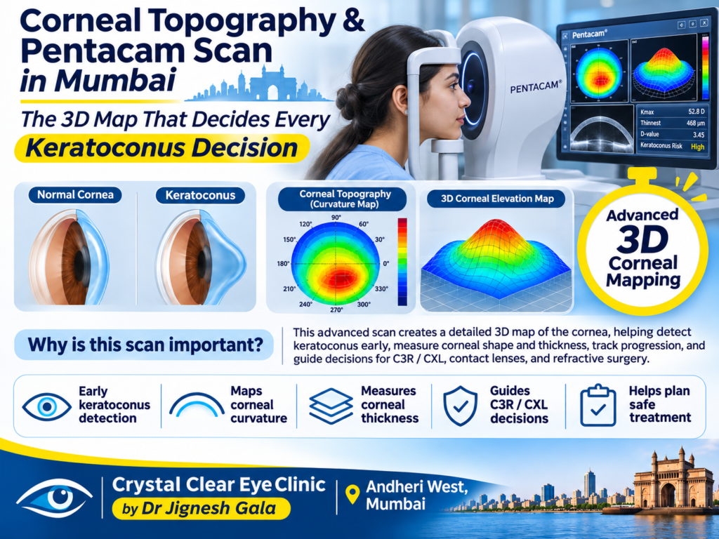
Corneal Topography & Pentacam Scan in Mumbai — The 3D Map That Decides Every Keratoconus Decision
Written & medically reviewed by Dr. Jignesh Gala, Vitreoretinal & Cataract Surgeon (FRCS Glasgow), Founder & Director — Crystal Clear Eye Clinic (Andheri West) and Topax Eye Hospital (Vile Parle East), Mumbai. Facts verified: 24 July 2026.
Thank you for reading this post, don't forget to subscribe!⚡ Quick answer: A corneal topography / Pentacam scan in Mumbai is a painless, 10-second, no-touch test that maps your cornea’s curvature, thickness and elevation in 3D — the single test that detects keratoconus years before symptoms, proves whether it is progressing (Kmax/ABCD grading), and decides between cross-linking, lenses, Intacs or monitoring. Cost: ₹1,200–2,500 (advanced tomography up to ₹5,000), verified 24 July 2026. Done on-site at Crystal Clear Eye Clinic, Andheri West and Topax Eye Hospital, Vile Parle East — and read by the doctor, not just printed. Call 077188 85245.
- What Is an Intravitreal Eye Injection?
- Drjigneshgalaeye.com
- A year at l v prasad eye institute how my vitreoretinal fellowship shaped the surgeon i am today
- About us
- Anti vegf injection cost in mumbai 2026 every drug every price explained honestly by a retina specialist
- B y l nair hospital 2019 2020 my sixteen months as assistant professor of ophthalmology
- Bakuchiol eye cream a gentle revolution in under eye care
- Best eye doctor for retina in mumbai dr jignesh gala retina vitreoretinal surgeon
- Best eye specialist in andheri mumbai
- Best retina specialist mumbai dr jignesh gala
- Best vision correction surgery after 40 what are your options
- Blogs by dr jignesh gala
1. What Corneal Topography/Tomography Is
Patients often use the two words interchangeably — here is the honest difference:
- Corneal topography is a curvature map — it measures the cornea’s front surface and paints it in colour codes (steep = red, flat = blue). Brilliant for astigmatism patterns and contact-lens work — but it only sees the front.
- Corneal tomography (the Pentacam-style Scheimpflug scan) goes further: a rotating camera photographs the cornea in cross-section slices and rebuilds a true 3D model — front surface, back surface, and thickness at every point (pachymetry). Keratoconus decisions are built on this test, because the earliest disease signs hide on the back surface and in the thickness map — places a front-only map cannot see.
The scan itself is almost comically easy: no drops, no touch, no injections — chin on a rest, look at a light, done in seconds per eye. Yet this ten-second scan carries more decision weight than any other test in keratoconus care — treatment timing, LASIK safety screening, progression proof. As our explainer on people do ask Google about this eye issue puts it: the test is cheap; the interpretation is the real product.
2. What the Scan Shows
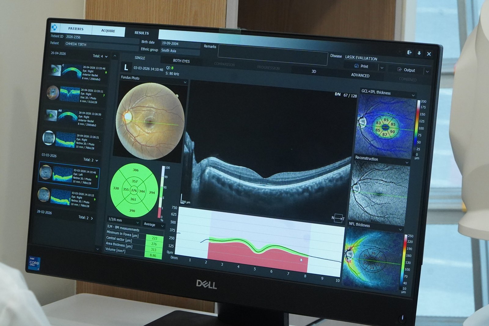
A Pentacam-style report is a wall of maps and numbers. Four matter most:
| Measurement | What It Is | Why It Matters in Keratoconus |
|---|---|---|
| K readings (K1, K2, Kmax) | Corneal curvature in dioptres; Kmax is the steepest point | Kmax is the number tracked over time — a rising Kmax means the cone is steepening (progression) |
| Pachymetry (thickness map) | Corneal thickness at every point, plus the thinnest location | Keratoconus is a thinning disease — thinnest-point values decide CXL safety and progression |
| Posterior elevation | How far the cornea’s back surface bulges from normal | The earliest keratoconus sign — abnormal years before routine exams show anything |
| ABCD staging indices | Composite grading from curvature, thickness and vision | Standardized severity/progression language used in serial comparison software |
Why this matters practically: a “normal” routine eye exam — even a normal front-surface topography — can coexist with early keratoconus; the back surface betrays the disease first. That is why we insist LASIK candidates (see RLE vs LASIK — which vision correction surgery is better and refractive lens exchange — the complete guide) get tomography, not a quick power check. One accuracy rule: RGP lens wearers need a 2-week washout before scanning, or the lens-moulded cornea gives false readings.
3. Progression Tracking — Why the Same Machine Matters
Diagnosing keratoconus once is easy. The question governing the rest of your care is harder: is it progressing? Progression is provable — but only with discipline:
- Serial comparison, not single scans. Belin-style analysis overlays your new scan on your baseline and flags change beyond test-to-test noise. We declare “progressing” only when Kmax, thinnest pachymetry and elevation move together beyond reproducibility limits — never on one nervous printout.
- Same machine, same centre. Machines measure differently; a Kmax of 48.2 on one system may read 47.6 on another with zero real change. Your 2026 scan must talk to your 2031 scan — possible only when the record lives in one place.
- Why it decides money and surgery. Proven progression triggers cross-linking; five-year registry data confirms CXL’s stabilizing effect (Save Sight registry study), and biomechanical studies confirm the cornea stays firmer years after (4-year longitudinal study). Stable scans, equally, are why we tell many patients not to spend on surgery.
Where cross-linking fits the map: when progression is proven, CXL halts it — it does not reverse the cone. Long-term outcomes are solid across protocols (accelerated vs standard riboflavin evidence, accelerated vs conventional results), biomaterials keep improving (2026 biomaterials review), and in selected cases topography-guided PRK with CXL improves vision while stabilizing (2024 cohort study). Every such decision starts from the scan.
4. Who Needs This Scan & How Often
| You Are… | Why You Need Tomography | How Often |
|---|---|---|
| A diagnosed keratoconus patient | Progression tracking — the basis of every treatment decision | Every 6–12 months on the same machine |
| A child/teen with keratoconus | Pediatric keratoconus progresses faster — earlier CXL saves corneas | Every 3–6 months |
| A LASIK/refractive surgery candidate | Mandatory safety screening — lasing an undetected cone is the classic refractive disaster | Once, before any procedure decision |
| Family member of a keratoconus patient | Keratoconus runs in families; early cones are silent | Baseline scan; repeat if abnormal |
| Eye-rubber / allergic patient with rising astigmatism | Frequent prescription changes + rubbing = screen the cornea | Once; repeat per findings |
| RGP/scleral lens wearer | Fit and corneal health monitoring (2-week RGP washout first) | With each follow-up as advised |
5. Topography/Pentacam Cost in Mumbai (2026)
Transparent, date-stamped numbers (verified 24 July 2026):
| Test | Mumbai 2026 Band | Notes |
|---|---|---|
| Corneal topography (front-surface map) | ₹1,200–2,500 | Useful for astigmatism patterns; not sufficient alone for keratoconus decisions |
| Corneal tomography / Pentacam scan | ₹1,200–2,500; advanced tomography up to ₹5,000 | The 3D gold standard — curvature + pachymetry + posterior elevation + ABCD indices |
| Serial progression analysis | Included in follow-up scans at our centres | New scan overlaid on your baseline — the report that actually answers “is it getting worse?” |
Honest guidance: the scan fee is not where value differs — every Mumbai Scheimpflug centre charges within this band. The value is in who reads it and whether it gets compared to your last one. A ₹1,500 scan filed away unread is money wasted; the same scan trended against your history is the foundation of every future decision. Call 077188 85245 for today’s exact price — a number, not a runaround. The same transparent approach drives our surgery packages page.
6. Why Patients Choose Dr. Jignesh Gala for Corneal Scanning

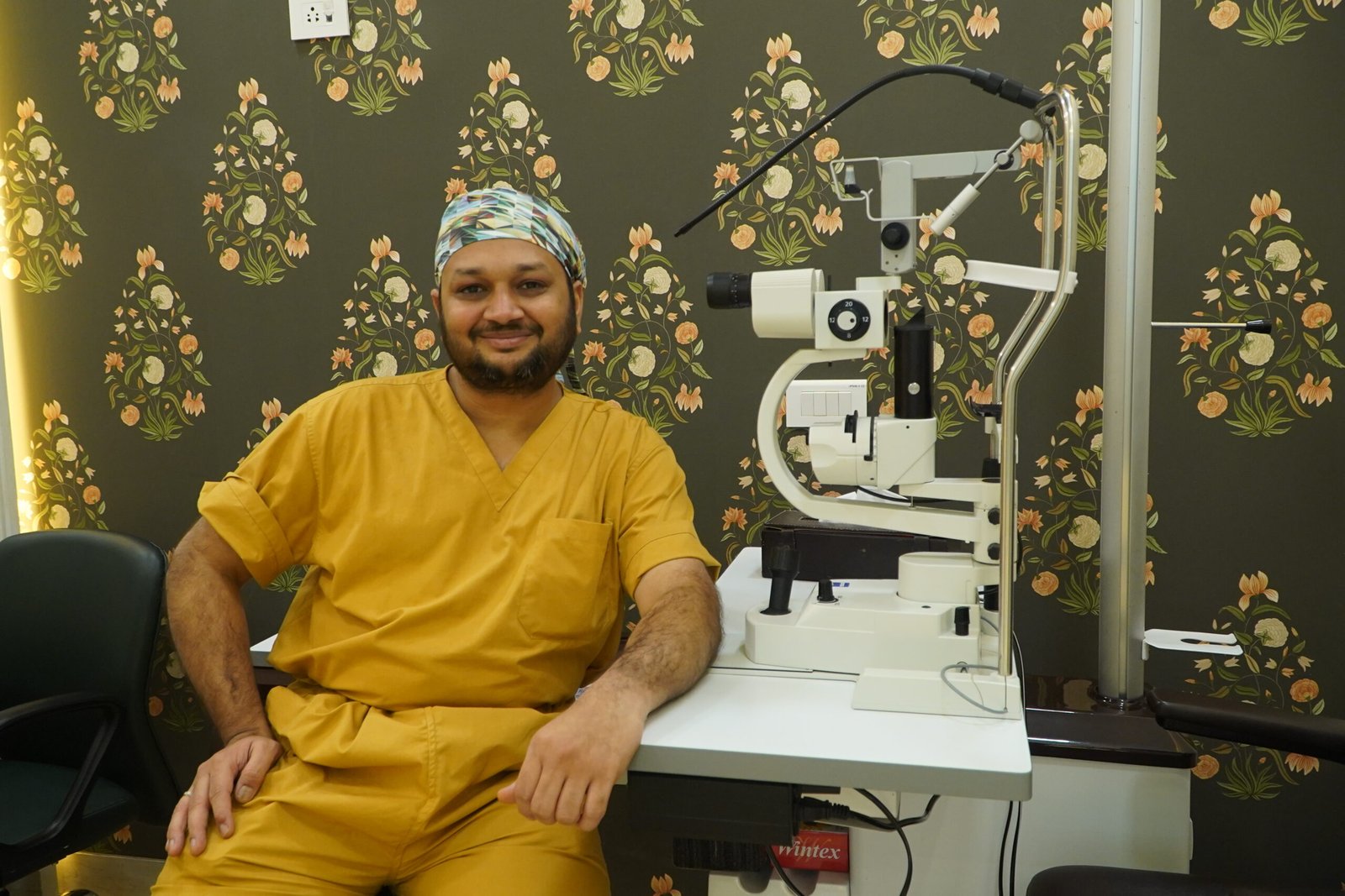
Dr. Jignesh Gala owns both Crystal Clear Eye Clinic (Andheri West) and Topax Eye Hospital (Vile Parle East) — your scan, your serial record, and whatever the scan leads to (cross-linking, lenses, Intacs planning) all live under one accountable roof.
✅ Doctor-read reports: Kmax, pachymetry maps, posterior elevation and ABCD indices explained on the screen, in Hindi, Marathi, Gujarati or English — never a technician handover.
✅ Trended, not printed-and-filed: every new scan compared against your baseline on the same machine; progression declared on evidence, not impression.
✅ The scan leads somewhere honest: proven progression → C3R/CXL at the centre; stable cone → monitoring and scleral-lens fitting with specialist fitters; lens-intolerant cones → Intacs evaluation; the ~10–15% truly needing DALK/PK → truthful counselling and planned referral with co-management, alternatives exhausted first.
✅ Research-current interpretation: fully aware of the latest research — unlike old practitioners who stopped reading journals years ago. He reads Cornea, JCRS, Journal of Refractive Surgery, AJO, BJO and IOVS, and spent six years as a BMJ Case Reports peer reviewer. His pedigree is public — refraction in real time — how the Royal College of Ophthalmologists tested me in Kuching 2017 and Singapore 2021 — a year on the founding ophthalmology team of Woodlands Health.
✅ Trusted beyond keratoconus: families who come for a Pentacam scan stay for everything — retina treatment in Andheri, advanced retinal care in Mumbai, retina laser treatment in Andheri West, retina surgery in Mumbai. See why in our satisfied patient stories.
7. Locations — Scans in Andheri West & Vile Parle East
1) Crystal Clear Eye Clinic — Andheri West
📍 Address: Laram Centre CHS, A1-202, Swami Vivekanand Road, above Sunil Jewellers, near NADCO Shopping Centre, Railway Colony, Andheri West, Mumbai 400058 • Open daily, closes 9 pm.
📞 Phone: 077188 85245
💳 Third-party/TPA cashless is done through Crystal Clear Eye Clinic (Andheri West).
Minutes from Lokhandwala, Versova, Juhu, Oshiwara, DN Nagar, Jogeshwari and Seven Bungalows; easy reach from Goregaon, Malad, Kandivali, Bandra, Khar and Santacruz. Searching corneal topography Pentacam scan Mumbai from the western suburbs ends here. Considering glass-removal surgery? Read why screening comes first in SmartSurf with CustomEyes — the most advanced glass removal surgery in Mumbai.

2) Topax Eye Hospital — Vile Parle East
📍 Vile Parle East, Mumbai • 📞 077188 85245
💳 Direct cashless is done through Topax Eye Care (Vile Parle East).
Convenient for Santacruz East, Khar, Bandra East, Kalina, Vakola, Juhu and the airport corridor. As a full hospital, Topax carries your scan forward into treatment — cross-linking, surgical planning, insurance-backed procedures — without your records ever leaving Dr. Gala’s system.
8. हिंदी • मराठी • ગુજરાતી — Patient Summaries
🇮🇳 हिंदी सारांश — मुंबई में कोर्नियल टोपोग्राफी / पेंटाकैम स्कैन
कोर्नियल टोपोग्राफी पेंटाकैम स्कैन मुंबई में यह 10-सेकंड की बेदर्द जांच आपकी कोर्निया का 3D नक्शा बनाती है — curvature, मोटाई (pachymetry) और posterior elevation। केराटोकोनस की पहचान और progression (Kmax/ABCD) का पूरा फैसला इसी स्कैन पर टिका है। कीमत ₹1,200–2,500 (verified 24 July 2026)। डॉ. गाला रिपोर्ट खुद पढ़कर समझाते हैं। अपॉइंटमेंट: 077188 85245।
🚩 मराठी सारांश — मुंबईत कॉर्नियल टोपोग्राफी / पेंटाकॅम स्कॅन
कॉर्नियल टोपोग्राफी पेंटाकॅम स्कॅन मुंबई मध्ये ही १०-सेकंदांची वेदनारहित तपासणी कॉर्नियाचा 3D नकाशा तयार करते — curvature, जाडी (pachymetry) व posterior elevation. केराटोकोनस ओळखणे व progression (Kmax/ABCD) ट्रॅक करणे यासाठीच हा स्कॅन आधार आहे. किंमत ₹1,200–2,500 (verified 24 July 2026). डॉ. गाला अहवाल स्वतः समजावतात. भेटीसाठी: 077188 85245.
🟡 ગુજરાતી સારાંશ — મુંબઈમાં કોર્નિયલ ટોપોગ્રાફી / પેન્ટાકેમ સ્કેન
કોર્નિયલ ટોપોગ્રાફી પેન્ટાકેમ સ્કેન મુંબઈમાં આ ૧૦-સેકન્ડની પીડારહિત તપાસ કોર્નિયાનો 3D નકશો બનાવે છે — curvature, જાડાઈ (pachymetry) અને posterior elevation. કેરાટોકોનસ ઓળખવો અને progression (Kmax/ABCD) ટ્રૅક કરવો — બધું આ જ સ્કેન પર આધારિત છે. કિંમત ₹1,200–2,500 (verified 24 July 2026). ડૉ. ગાલા રિપોર્ટ જાતે સમજાવે છે. એપોઈન્ટમેન્ટ: 077188 85245.
9. ❓ 8 Frequently Asked Questions (English + Hinglish)
1. Corneal topography kya hoti hai?
Cornea ka colour-coded curvature map (steep = red, flat = blue). Uski advanced behen tomography (Pentacam) hai — poori cornea ka 3D model: aage ki surface, peechhe ki surface, har point ki motai. Keratoconus decisions ke liye tomography zaroori hai, kyunki shuruaati signs peechhe ki surface par hote hain.
2. Pentacam scan cost in Mumbai kitna hai?
Corneal tomography/Pentacam ₹1,200–2,500; advanced tomography up to ₹5,000 (verified 24 July 2026). Exact price: call 077188 85245. Yaad rakhein — value scan fee mein nahi, interpretation aur serial comparison mein hai.
3. Kya Pentacam scan painful hai?
Bilkul nahi. Koi drops nahi, koi touch nahi, koi injection nahi. Chin rest par rakhiye, ek light ki taraf dekhiye — machine seconds mein scan complete kar leti hai. Bachche bhi aaraam se karwa lete hain; report turant ready.
4. How often should keratoconus patients repeat tomography?
Every 6–12 months for stable adults; every 3–6 months for children and teens (pediatric keratoconus progresses faster) and after cross-linking. Always on the same machine — only then can the new scan be trended against your baseline.
5. Topography aur tomography mein kya farak hai?
Topography sirf cornea ki front surface ka curvature map hai. Tomography (Pentacam/Scheimpflug) poori cornea ka 3D model banati hai — front + back surface + thickness maps + posterior elevation. Early keratoconus signs back surface aur thinning mein hote hain, isliye diagnosis aur progression tracking ke liye tomography chahiye.
6. Does LASIK need a Pentacam scan first?
Yes — mandatory. Lasing an undetected early cone is the classic refractive-surgery disaster (post-LASIK ectasia). Every responsible LASIK evaluation includes tomography to rule out keratoconus. If a LASIK centre skips tomography, that is your answer about that centre.
7. Corneal topography kahan hoti hai Andheri West mein?
Crystal Clear Eye Clinic — Laram Centre CHS, A1-202, S.V. Road, above Sunil Jewellers, near NADCO Shopping Centre, Andheri West 400058; ya Topax Eye Hospital, Vile Parle East. Phone: 077188 85245. Open daily, closes 9 pm. Same-day scan, doctor-read report, serial record humare paas hi rehta hai.
8. Report kaun padhta hai — technician ya doctor?
Bahut se centres mein technician scan karke printout de deta hai. Humare yahan Dr. Gala khud har report padhte hain — Kmax, thinnest pachymetry, posterior elevation, ABCD — aur screen par aapki bhasha mein samjhaate hain. Scan machine karta hai; faisla doctor ka hota hai.
13. 📖 Related Reading — Complete Eye-Care Library by Dr. Jignesh Gala (78 guides)
Every guide below is written by the same surgeon who would operate on you:
From drjigneshgalaeye.com
- Drjigneshgalaeye.com
- A year at l v prasad eye institute how my vitreoretinal fellowship shaped the surgeon i am today
- About us
- Anti vegf injection cost in mumbai 2026 every drug every price explained honestly by a retina specialist
- B y l nair hospital 2019 2020 my sixteen months as assistant professor of ophthalmology
- Bakuchiol eye cream a gentle revolution in under eye care
- Best eye doctor for retina in mumbai dr jignesh gala retina vitreoretinal surgeon
- Best eye specialist in andheri mumbai
- Best retina specialist mumbai dr jignesh gala
- Best vision correction surgery after 40 what are your options
- Blogs by dr jignesh gala
- Cashless eye surgery in mumbai dr jignesh gala
- Diabetic retinopathy specialist in andheri west mumbai advanced screening injections laser vitrectomy by dr jignesh gala frcs
- Discover crystal clear vision with contoura lasik in mumbai
- Dr jignesh gala top eye specialist in mumbai
- Drjignesh gala premier pediatric ophthalmologistandheri mumbai
- Eylea injection in mumbai cost uses side effects the 2026 price revolution explained by a retina specialist
- Glaucoma treatment in andheri mumbai
- How to prevent dry eyes in winter
- Hvf test in andheri west mumbai visual field test cost procedure how to read your report by a specialist
- I see a curtain or shadow in my vision
- Inside my six years as a bmj case reports peer reviewer
- Inside the fico 2016 the only worldwide medical specialty examination
- Inside the frcs ophth glasgow how a 425 year old royal college tests your surgical knowledge
- Intravitreal injection in andheri west mumbai painless anti vegf eye injections by dr jignesh gala frcs
- Know your eye doctor
- Laser cataract surgery in andheri mumbai dr jignesh gala
- Laser cataract surgery
- Lasik eye surgery in andheri mumbai dr jignesh gala
- Lasik eye surgery in bandra mumbai dr jignesh gala
- Lasik eye surgery in juhu dr jignesh gala
- Lasik eye surgery in lokhandwala dr jignesh gala
- Lasik eye surgery in oshiwara dr jignesh gala
- Lasik in santacruz mumbai
- Lasik in versova mumbai dr jignesh gala
- Lasik surgery in andheri mumbai
- Most common types of refractive eye surgery which one is right for you
- Mrcs edinburgh how i joined a royal college founded in 1505
- Multifocal trifocal iol guide complete patient guide
- Oct eye test in andheri west mumbai cost procedure report why the specialist reading it matters
- People do ask google about this eye issue
- Refraction in real time how the royal college of ophthalmologists tested me in kuching 2017
- Refractive lens exchange rlethe complete guide to clear vision without glasses
- Retina laser treatment in andheri west mumbai prp barrage focal micropulse laser by dr jignesh gala frcs
- Retina surgery in mumbai your complete guide to saving your sight
- Retina treatment in andheri
- Retina treatment in mumbai advanced retinal care by dr jignesh gala
- Rle vs lasik which vision correction surgery is better
- Satisfied patient stories
- Singapore 2021 a year on the founding ophthalmology team of woodlands health
- Smartsurf with customeyes the most advanced glass removal surgery in mumbai and why it outperforms wavelight plus innoveyes
- Surgery packages
- Surgical videos by dr jignesh gala
- Types of intraocular lenses
- Types of refractive surgeries
- Types of retina surgeries
- Understanding and preventing digital eye strain
From crystalcleareye.in
- Crystal Clear Eye Clinic — Home
- About Crystal Clear Eye Clinic, Andheri West
- Eye Health Blog — Crystal Clear Eye Clinic
- Contact & Directions — Andheri West
- Glaucoma Treatment in Andheri, Mumbai
- LASIK Surgery in Andheri, Mumbai
- Premium IOL Selection Guide 2026
- Laser Cataract Surgery in Andheri, Mumbai
- Best Eye Specialist in Andheri, Mumbai
- Best Retina Specialist in Mumbai
- Best Vitrectomy Surgery in Mumbai — Expert Retinal Care
- Diabetic Eye Care in Mumbai
- Diabetic Retinopathy in Andheri
- Diabetic Macular Edema (DME) Treatment in Mumbai
- Macular Edema Treatment in Andheri West
- ARMD Treatment in Andheri West, Mumbai
- Retina Treatment in Andheri
- Vitreoretinal Treatment in Andheri
- Cashless Eye Surgery in Mumbai
- Sudden Floaters? Retina Check-up in Mumbai
- What Is an Intravitreal Eye Injection?
Your retina deserves a specialist. Today, not someday.
Every keratoconus decision — watch, cross-link, lenses, surgery — begins with one 3D map of your cornea.
📍 Andheri West: Laram Centre CHS, A1-202, S.V. Road, above Sunil Jewellers, near NADCO Shopping Centre, Mumbai 400058
📍 Vile Parle East: Topax Eye Hospital • Open daily, closes 9 pm
Dr. Jignesh Gala, FRCS (Glasgow) — Vitreoretinal & Cataract Surgeon • drjigneshgalaeye.com • crystalcleareye.in
Medical disclaimer: This page is educational and does not replace a personal consultation. Prices are indicative, verified on 24 July 2026, and may change. Every eye is different — decisions are made only after examination by Dr. Jignesh Gala.
Research & Evidence Library — 110 Verified References
Verified live in July 2026 via official NCBI E-utilities — including research from Cornea, JCRS, Journal of Refractive Surgery, AJO, BJO, IOVS, Eye & Contact Lens, IJO, Acta Ophthalmologica, Graefe’s Archive and TVST. The same journals Dr. Gala reads. Click any title to check the science yourself.
KC: Corneal Tomography / Pentacam Diagnosis & Progression
- Corneoscleral Morphology in Keratoconus and Its Association With Corneal Tomography and Biomechanics. — Translational vision science & technology, 2026
- Simultaneous Topography-guided Custom Ablation With Corneal Cross-linking for Keratoconus: 10-Year Prospective Outcomes. — Journal of refractive surgery (Thorofare, N.J. : 1995), 2023
- Superiority of Baseline Biomechanical Properties over Corneal Tomography in Predicting Keratoconus Progression. — Turkish journal of ophthalmology, 2021
- Machine Learning-Based Identification of Risk Factors of Keratoconus Progression Using Raw Corneal Tomography Data. — Cornea, 2024
- Topical Losartan for Treating Corneal Haze After Ultraviolet-A/Riboflavin Collagen Cross-Linking. — Cornea, 2024
- Repeatability and Comparison of Corneal Tomography in Mild to Severe Keratoconus Between the Anterior Segment OCT MS-39 and Pentacam HR. — Journal of refractive surgery (Thorofare, N.J. : 1995), 2022
- Keratoconus tomographic indices in osteogenesis imperfecta. — Graefe’s archive for clinical and experimental ophthalmology = Albrecht von Graefes Archiv fur klinische und experimentelle Ophthalmologie, 2023
- Belin ABCD Progression Display Identifies Keratoconus Progression Earlier Than Conventional Metrics. — American journal of ophthalmology, 2022
- Scheimpflug-Derived Keratometric, Pachymetric and Pachymetric Progression Indices in the Diagnosis of Keratoconus: A Systematic Review and Meta-Analysis. — Clinical ophthalmology (Auckland, N.Z.), 2023
- Corneal cross-linking in patients with keratoconus: up to 13 years of follow-up. — Graefe’s archive for clinical and experimental ophthalmology = Albrecht von Graefes Archiv fur klinische und experimentelle Ophthalmologie, 2023
- Influence of Keratoconus Severity on Detecting True Progression with Scheimpflug Imaging and Anterior Segment Optical Coherence Tomography. — Life (Basel, Switzerland), 2023
- Artificial intelligence for detecting keratoconus. — The Cochrane database of systematic reviews, 2023
- Enhancing early detection of keratoconus suspects using interocular corneal tomography asymmetry. — International ophthalmology, 2025
- Early diagnosis of keratoconus using corneal biomechanics and OCT derived technologies. — Eye and vision (London, England), 2025
- Analysis of Scheimpflug Tomography Parameters for Detecting Subclinical Keratoconus in the Fellow Eyes of Patients with Unilateral Keratoconus in the Eastern Province of Saudi Arabia. — Clinical ophthalmology (Auckland, N.Z.), 2024
KC: Artificial Intelligence Detection (Tomography/Biomechanics)
- Enhancing Early Keratoconus Detection With Multimodal Machine Learning: Integrating Tomography, Biomechanics, and Clinical Risk Factors. — American journal of ophthalmology, 2025
- Keratoconus: imaging modalities and management. — Medical hypothesis, discovery & innovation ophthalmology journal, 2024
- [Detection of Subclinical Keratoconus]. — Klinische Monatsblatter fur Augenheilkunde, 2022
- Advancing subclinical keratoconus detection using polarization-sensitive optical coherence tomography and artificial intelligence. — Biophotonics discovery, 2026
- Optimized Artificial Intelligence for Enhanced Ectasia Detection Using Scheimpflug-Based Corneal Tomography and Biomechanical Data. — American journal of ophthalmology, 2023
- Artificial Intelligence-Derived Biomechanical Index for Early Corneal Ectasia Detection: Advancing Beyond Tomography. — Ophthalmology science, 2026
- Early Detection of Keratoconus: Diagnostic Advances and Their Impact on Visual Outcomes: A Systematic Review. — Medicina (Kaunas, Lithuania), 2025
- Artificial intelligence in ophthalmology: opportunities, challenges, and ethical considerations. — Medical hypothesis, discovery & innovation ophthalmology journal, 2025
- A Novel Chaos-inspired Artificial Intelligence-based Model for Keratoconus Prediction. — Journal of ophthalmic & vision research, 2026
- Risk factors for postlaser refractive surgery corneal ectasia. — Current opinion in ophthalmology, 2020
- Machine Learning Algorithms to Detect Subclinical Keratoconus: Systematic Review. — JMIR medical informatics, 2021
- Multimodal diagnostics for keratoconus and ectatic corneal diseases: a paradigm shift. — Eye and vision (London, England), 2023
- Update on topography and tomography for refractive surgery. — Current opinion in ophthalmology, 2026
- Enhanced Diagnostics for Corneal Ectatic Diseases: The Whats, the Whys, and the Hows. — Diagnostics (Basel, Switzerland), 2022
- Role of corneal epithelial mapping, Corvis biomechanical index, and artificial intelligence-based tomographic biomechanical index in diagnosing spectrum of keratoconus. — Oman journal of ophthalmology, 2023
OCT: Diabetic Macular Edema Biomarkers
- Multimodal imaging in diabetic retinopathy and macular edema: An update about biomarkers — Survey of ophthalmology, 2024
- Persistent diabetic macular edema: Definition, incidence, biomarkers, and treatment methods — Survey of ophthalmology, 2023
- Baseline OCT Biomarkers Associated with Visual Acuity in Diabetic Macular Edema: A Systematic Review and Meta-analysis — Ophthalmology, 2026
- Optical coherence tomography in the management of diabetic macular oedema — Progress in retinal and eye research, 2024
- Imaging and Biomarkers in Diabetic Macular Edema and Diabetic Retinopathy — Current diabetes reports, 2019
OCT: Oct Angiography In Diabetic Retinopathy
- OCT angiography 2023 update: focus on diabetic retinopathy — Acta diabetologica, 2024
- New insights into diabetic retinopathy by OCT angiography — Diabetes research and clinical practice, 2018
- Optical coherence tomography angiography in diabetic retinopathy: an updated review — Eye (London, England), 2021
- Association of OCT Angiography-Detected Intraretinal Microvascular Abnormalities with Diabetic Retinopathy Severity — Ophthalmology. Retina, 2026
- COMPARATIVE ANALYSIS OF OCT AND OCT ANGIOGRAPHY CHARACTERISTICS IN EARLY DIABETIC RETINOPATHY — Retina (Philadelphia, Pa.), 2023
OCT: Glaucoma – Retinal Nerve Fiber Layer
- Retinal Nerve Fiber Layer Optical Texture Analysis: Involvement of the Papillomacular Bundle and Papillofoveal Bundle in Early Glaucoma — Ophthalmology, 2022
- Anatomical compensation of retinal nerve fiber layer improves the detection of glaucoma between ethnicities — Annals of the New York Academy of Sciences, 2024
- Racial Differences in Diagnostic Accuracy of Retinal Nerve Fiber Layer Thickness in Primary Open-Angle Glaucoma — American journal of ophthalmology, 2024
- Peripapillary Retinal Nerve Fiber Layer Vascular Microcirculation in Glaucoma Using Optical Coherence Tomography-Based Microangiography — Investigative ophthalmology & visual science, 2016
- Evaluating the retinal nerve fiber layer in glaucoma with scanning laser polarimetry — Archives of ophthalmology (Chicago, Ill. : 1960), 1999
OCT: Telemedicine / Screening For Diabetic Retinopathy
- Review of smartphone funduscopy for diabetic retinopathy screening — Survey of ophthalmology, 2024
- Comparison of Telemedicine Screening of Diabetic Retinopathy by Mydriatic Smartphone-Based vs Nonmydriatic Tabletop Camera-Based Fundus Imaging — Journal of vitreoretinal diseases, 2021
- The role of tele-ophthalmology in diabetic retinopathy screening — Journal of optometry, 2020
- Cost-effectiveness of a National Telemedicine Diabetic Retinopathy Screening Program in Singapore — Ophthalmology, 2016
- Feasibility of telemedicine in detecting diabetic retinopathy and age-related macular degeneration — Seminars in ophthalmology, 2015
OCT: Artificial Intelligence In Oct Interpretation
- [OCT biomarkers in diabetic maculopathy and artificial intelligence] — Die Ophthalmologie, 2024
- Artificial intelligence for retinal diseases — Asia-Pacific journal of ophthalmology (Philadelphia, Pa.), 2024
- Artificial Intelligence and Imaging Processing in Optical Coherence Tomography and Digital Images in Uveitis — Ocular immunology and inflammation, 2022
- Application of Artificial Intelligence in Targeting Retinal Diseases — Current drug targets, 2020
- A comparison of deep learning performance against health-care professionals in detecting diseases from medical imaging: a systematic review and meta-analysis — The Lancet. Digital health, 2019
OCT: Macular Hole & Epiretinal Membrane
- Epiretinal membrane: an overview and update — Japanese journal of ophthalmology, 2024
- In-Depth Analysis of Preoperative OCT Markers as Prognostic Factors for Lamellar Macular Holes and Epiretinal Membrane Foveoschisis — Ophthalmology. Retina, 2024
- Macular hole with epiretinal proliferation: diagnostic value of en-face optical coherence tomography and clinical characteristics — Graefe’s archive for clinical and experimental ophthalmology = Albrecht von Graefes Archiv fur klinische und experimentelle Ophthalmologie, 2024
- The clinical and pathogenic significance of atypical epiretinal tissue in macular hole — Graefe’s archive for clinical and experimental ophthalmology = Albrecht von Graefes Archiv fur klinische und experimentelle Ophthalmologie, 2022
- Optical coherence tomography in the preoperative and postoperative management of macular hole and epiretinal membrane — The British journal of ophthalmology, 2014
OCT: Central Serous Chorioretinopathy
- Central serous chorioretinopathy and the sclera: what we have learned so far — Japanese journal of ophthalmology, 2024
- Can we classify central serous chorioretinopathy better? Yes we can — Eye (London, England), 2022
- Diagnostic accuracy of OCT angiography for macular neovascularization in central serous chorioretinopathy: A systematic review and meta-analysis — Acta ophthalmologica, 2024
- Regression patterns of central serous chorioretinopathy using en face optical coherence tomography — Graefe’s archive for clinical and experimental ophthalmology = Albrecht von Graefes Archiv fur klinische und experimentelle Ophthalmologie, 2022
- Central Serous Chorioretinopathy Analyzed by Multimodal Imaging — Translational vision science & technology, 2021
OCT: Age-Related Macular Degeneration Biomarkers
- OCT Prognostic Biomarkers for Progression to Late Age-related Macular Degeneration: A Systematic Review and Meta-analysis — Ophthalmology. Retina, 2024
- Baseline OCT Biomarkers Predicting Visual Outcomes in Neovascular Age-Related Macular Degeneration: A Meta-Analysis — Ophthalmology, 2025
- Choroidal biomarkers in age-related macular degeneration — Survey of ophthalmology, 2025
- Practical guidance for imaging biomarkers in exudative age-related macular degeneration — Survey of ophthalmology, 2023
- Predicting Progression of Age-Related Macular Degeneration Using OCT and Fundus Photography — Ophthalmology. Retina, 2021
OCT: Vitreomacular Traction
- Vitreomacular traction – a review — Eye (London, England), 2025
- Vitreomacular traction in diabetic retinopathy — Japanese journal of ophthalmology, 2024
- Vitreomacular Traction Surgery from the DISCOVER Study: Intraoperative OCT Utility, Ellipsoid Zone Dynamics, and Outcomes — Ophthalmic surgery, lasers & imaging retina, 2021
- A Study of the Natural History of Vitreomacular Traction Syndrome by OCT — Ophthalmology, 2018
- The International Vitreomacular Traction Study Group classification of vitreomacular adhesion, traction, and macular hole — Ophthalmology, 2013
OCT: Swept-Source Oct / Enhanced Depth Imaging Choroid
- Choroidal biomarkers — Indian journal of ophthalmology, 2018
- Enhanced depth imaging in swept-source optical coherence tomography: Improving visibility of choroid and sclera, a masked study — European journal of ophthalmology, 2020
- Choroidal imaging biomarkers — Survey of ophthalmology, 2019
- Enhanced depth imaging-OCT of the choroid: a review of the current literature — Graefe’s archive for clinical and experimental ophthalmology = Albrecht von Graefes Archiv fur klinische und experimentelle Ophthalmologie, 2014
- Enhanced depth imaging optical coherence tomography — Ophthalmic surgery, lasers & imaging : the official journal of the International Society for Imaging in the Eye, 2011
KC: Accelerated Vs Conventional Cross-Linking (Epi-On)
- Keratoconus Treatment Algorithm. — Ophthalmology and therapy, 2017
- Expanding indications for corneal cross-linking. — Current opinion in ophthalmology, 2023
- Accelerated Epi-On Versus Standard Epi-Off Corneal Collagen Cross-Linking for Progressive Keratoconus in Pediatric Patients: Five Years of Follow-Up. — Cornea, 2020
- Accelerated Epi-On Versus Standard Epi-Off Corneal Collagen Cross-Linking for Progressive Keratoconus in Pediatric Patients. — Cornea, 2017
- A Transepithelial Corneal Cross-linking (CXL) Protocol Providing the Same Biomechanical Strengthening as Accelerated Epithelium-off CXL. — Journal of refractive surgery (Thorofare, N.J. : 1995), 2025
- A Review of Keratoconus Cross-Linking Treatment Methods. — Journal of clinical medicine, 2025
- Accelerated epithelium-on or accelerated epithelium-off corneal collagen cross-linking: Contralateral comparison study. — Taiwan journal of ophthalmology, 2020
- Progressive high-fluence epithelium-on accelerated corneal crosslinking: a novel corneal photodynamic therapy for early progressive keratoconus. — Frontiers in medicine, 2023
- Efficacy and safety of progressive fluence pulsed light epithelium-on accelerated corneal cross-linking for progressive keratoconus: 18-month prospective results. — International journal of ophthalmology, 2026
- Visual and Topographic Improvement with Epithelium-On, Oxygen-Supplemented, Customized Corneal Cross-Linking for Progressive Keratoconus. — Journal of clinical medicine, 2020
- Optimization of Oxygen Dynamics, UV-A Delivery, and Drug Formulation for Accelerated Epi-On Corneal Crosslinking. — Current eye research, 2020
- Epithelium-on Corneal Cross-linking Using a Modified Penetration Enhancer Without Supplemental Oxygen or Iontophoresis. — Journal of refractive surgery (Thorofare, N.J. : 1995), 2026
- Topo-Pachimetric Accelerated Epi-On Cross-Linking Compared to the Dresden Protocol Using Riboflavin with Vitamin E TPGS: Results of a 2-Year Randomized Study. — Journal of clinical medicine, 2021
- Management of Tomographically Suspicious Fellow Eyes of Young Patients With Unilateral Clinically Evident Keratoconus: Accelerated Epithelium-On Corneal Crosslinking Versus Observation. — Cornea, 2025
- Sterile corneal infiltrates after corneal collagen cross-linking: evaluation of risk factors. — Acta ophthalmologica, 2017
KC: Scleral Lenses: Visual Rehabilitation
- Keratoconus Diagnosis and Treatment: Recent Advances and Future Directions. — Clinical ophthalmology (Auckland, N.Z.), 2023
- Visual rehabilitation with mini scleral contact lenses in scarred corneas. — Journal francais d’ophtalmologie, 2024
- Efficacy of mini-scleral lenses in visual rehabilitation for corneal disorders: a prospective study. — BMC ophthalmology, 2025
- Visual rehabilitation using SPOT(®) scleral lenses in patients with keratoconus. — European journal of ophthalmology, 2026
- Visual rehabilitation using mini-scleral contact lenses after penetrating keratoplasty. — Arquivos brasileiros de oftalmologia, 2017
- Refractive approaches to visual rehabilitation in patients with keratoconus. — Current opinion in ophthalmology, 2020
- Visual Rehabilitation of Bilateral Keratoconus With Customized Scleral Lenses: A Case Report. — Cureus, 2026
- Safety and Efficacy of Scleral Lenses for Keratoconus. — Optometry and vision science : official publication of the American Academy of Optometry, 2020
- Contact lenses for visual rehabilitation in post-keratoplasty eyes: A review. — Indian journal of ophthalmology, 2025
- Scleral contact lenses for visual rehabilitation after penetrating keratoplasty: long term outcomes. — Contact lens & anterior eye : the journal of the British Contact Lens Association, 2014
- Mini-Scleral Lens: An Effective Mode of Visual Rehabilitation in Eyes With Resolved Hydrops. — Eye & contact lens, 2024
- Influence of Scleral Contact Lenses on Optical Coherence Tomography Parameters in Keratoconus Patients. — Diagnostics (Basel, Switzerland), 2025
- Impact of Scleral Lenses on Visual Acuity and Ocular Aberrations in Corneal Ectasia: A Comprehensive Review. — Journal of personalized medicine, 2024
- Irregular Corneas: Improve Visual Function With Scleral Contact Lenses. — Eye & contact lens, 2018
- Visual performance of scleral lenses and their impact on quality of life in patients with irregular corneas. — Arquivos brasileiros de oftalmologia, 2018