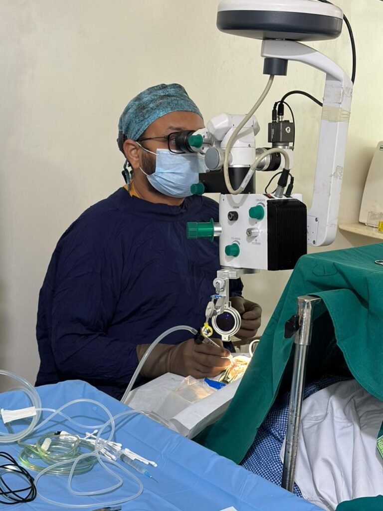
Top Retina Surgeon & Specialist in Mumbai — Why Dr. Jignesh Gala Is Trusted With the City’s Most Complex Eyes
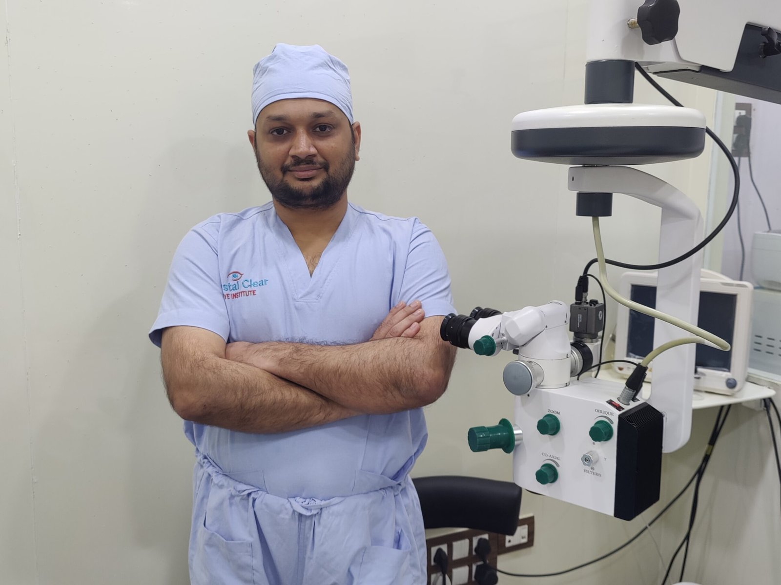
Written & medically reviewed by Dr. Jignesh Gala, Vitreoretinal & Cataract Surgeon (FRCS Glasgow), Founder & Director — Crystal Clear Eye Clinic (Andheri West) and Topax Eye Hospital (Vile Parle East), Mumbai. Facts verified: 24 July 2026.
Thank you for reading this post, don't forget to subscribe!🚨 Retinal emergency? A sudden curtain, shadow or storm of floaters means the clock is running. Call 077188 85245 now — seen same-day at Crystal Clear Eye Clinic, Andheri West. Open daily, closes 9 pm.
⚡ Quick answer: The best retina surgeon in Mumbai is not the one with the biggest billboard — it’s the one with subspecialty fellowship training, proven complex-case volume, current research practice, and hospitals of his own. Dr. Jignesh Gala — FRCS (Glasgow), MRCS (Edinburgh), FICO, vitreoretinal fellow at L.V. Prasad Eye Institute, former Assistant Professor at B.Y.L. Nair Hospital, founding ophthalmology team at Woodlands Health Singapore, six-year BMJ Case Reports peer reviewer — operates the full range of complex retinal problems at his own centres, Crystal Clear Eye Clinic (Andheri West) and Topax Eye Hospital (Vile Parle East). Consult him directly on 077188 85245.
- Lasik in versova mumbai dr jignesh gala
- Lasik surgery in andheri mumbai
- Most common types of refractive eye surgery which one is right for you
- Mrcs edinburgh how i joined a royal college founded in 1505
- Multifocal trifocal iol guide complete patient guide
- Oct eye test in andheri west mumbai cost procedure report why the specialist reading it matters
- People do ask google about this eye issue
- Refraction in real time how the royal college of ophthalmologists tested me in kuching 2017
- Refractive lens exchange rlethe complete guide to clear vision without glasses
- Retina laser treatment in andheri west mumbai prp barrage focal micropulse laser by dr jignesh gala frcs
- Retina surgery in mumbai your complete guide to saving your sight
- Retina treatment in andheri
1. What “Top Retina Surgeon” Actually Means — The Verification Checklist
Type “best retina surgeon in Mumbai” or “top retina specialist in Mumbai” into Google and a hundred names appear. Most lists are paid placements. Here is the checklist that actually separates a world-class vitreoretinal surgeon from a general eye doctor:
- Fellowship, not just degree. An MS/DNB ophthalmologist is a generalist. A vitreoretinal fellowship is 1–2 years operating only on the back of the eye, under masters. Ask where it was done.
- International examination credentials. FRCS (Glasgow), MRCS (Edinburgh), FICO — these are not memberships bought online; they are exit examinations passed abroad.
- Complex-case volume. A surgeon who sees routine cataracts all day is not the one for your PVR detachment. Ask: “Do you take referred and failed cases?”
- His own operation theatre. The surgeon who owns his OT controls the machines, the disposables, the sterility and the scheduling — and is there on a Sunday when your retina tears.
- Research currency. Retina surgery changed more between 2016 and 2026 than in the previous forty years. If your surgeon stopped reading journals, his technique stopped evolving.
- Honest, written outcomes. Real specialists quote ranges and risks, not “100% success.”
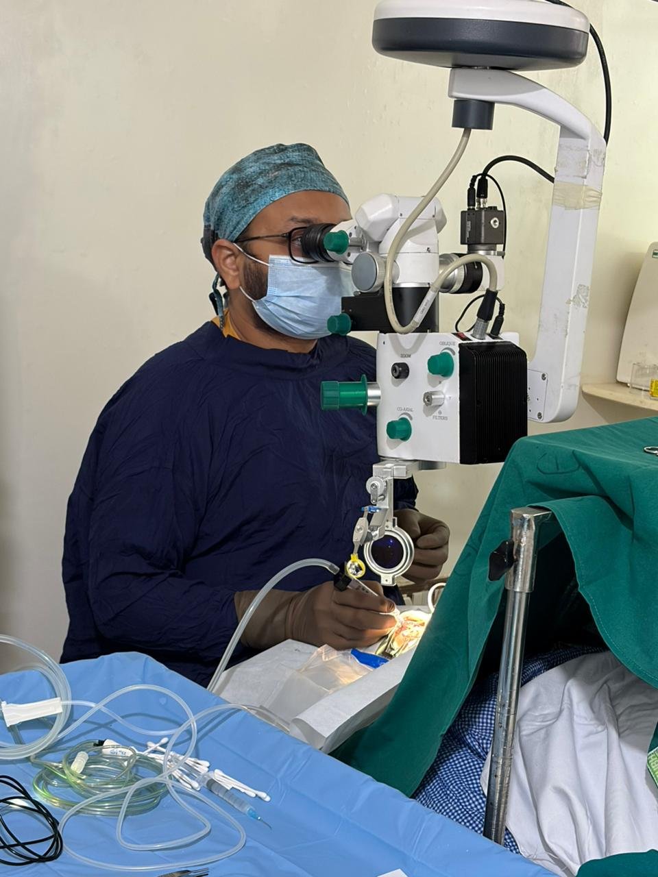
2. Dr. Jignesh Gala Against the Checklist — Point by Point
| Checklist Item | Dr. Gala’s Proof |
|---|---|
| Subspecialty fellowship | Vitreoretinal fellowship, L.V. Prasad Eye Institute — Asia’s premier retina institute, where complicated cases are the daily diet |
| International exams | FRCS (Glasgow), MRCS (Edinburgh), FICO — the complete international credential set |
| Academic depth | Former Assistant Professor of Ophthalmology, B.Y.L. Nair Hospital, Mumbai — a municipal-hospital tenure built on trauma, infection and referred failures |
| Global exposure | Founding ophthalmology team, Woodlands Health, Singapore — top-class training across India, UK exams and Singapore |
| Peer-trusted judgment | Six years as a BMJ Case Reports peer reviewer — an international journal asks him to judge other doctors’ complex cases |
| Own hospitals | Owns both Crystal Clear Eye Clinic (Andheri West) and Topax Eye Hospital (Vile Parle East) — no visiting-surgeon handoffs |
| Complex cases | PVR detachments, diabetic traction, dropped nucleus rescue, scleral-fixated IOL, endophthalmitis, open-globe trauma, failed surgeries referred from elsewhere |
3. The Complex Retinal Problems We Solve Every Week
The cases other clinics call “too difficult” are the cases this clinic was built for:
- Retinal detachment — including PVR and re-detachments. Micro-incision 23/25-gauge vitrectomy with gas or silicone oil, scleral buckle where it serves better, pneumatic retinopexy for selected cases. Primary success 90–95% (2024 review).
- Diabetic tractional detachment and vitreous hemorrhage. Pre-op anti-VEGF, meticulous membrane delamination, endolaser — the hardest surgery in ophthalmology, done routinely (diabetic TRD outcomes).
- Macular hole — including large, chronic and failed holes. ILM peel with inverted-flap technique, the closure method with the strongest evidence (inverted flap vs ILM peel).
- Dropped nucleus and dislocated IOL. Complete vitrectomy, fragment removal, secondary scleral-fixated lens (sutured, glued or Yamane) — finished in one admission.
- Endophthalmitis. The 48-hour emergency: tap, antibiotics, early vitrectomy when indicated — following the latest timing evidence (when to vitrectomise).
- Failed or referred surgeries. Recurrent detachments, persistent holes, retained lens material — a proper second surgical opinion with a written rescue plan.
- The whole routine retina spectrum too: diabetic retinopathy screening and injections, ARMD (wet AMD) management, retinal vein occlusion, epiretinal membranes, myopic degeneration, retinal tears and barrage laser.
4. Research-Current Surgery: What 2026 Demands
Dr. Gala is fully aware of the latest research — unlike old practitioners who stopped reading journals years ago. What that means in the operating theatre and the injection room:
- Smaller wounds, faster healing: 25-gauge micro-incision platforms as the default, not the exception.
- Drug-era retina care: the 2026 anti-VEGF landscape (Eylea’s price revolution, biosimilars, faricimab) changes when and how often eyes need surgery at all.
- Evidence before ego: PIVOT-trial-era choices between pneumatic retinopexy and vitrectomy; inverted-flap versus simple ILM peel; the re-examined endophthalmitis guidelines — each technique on this page cites its evidence (library below).
- Diagnostics that decide: OCT and OCTA read personally by the surgeon, visual fields trended, progression measured before any knife is lifted.
5. Honest Outcomes & Transparent Costs
| Procedure | Typical Success (Expert Hands) | Mumbai Cost Band (2026) |
|---|---|---|
| Primary retinal detachment repair | 90–95% single-surgery reattachment | ₹50,000–1,50,000 |
| Macular hole closure | 90–95% (85–90% large/chronic) | ₹45,000–1,20,000 |
| Diabetic tractional detachment | 85–90% anatomical success | ₹70,000–1,50,000 |
| Dropped-nucleus rescue + secondary IOL | >90% completed in one admission | ~₹70,000 (SFIOL benchmark) |
| Anti-VEGF injections | Disease-specific; vision stabilization is the goal | ₹8,000–38,000 per injection (Eylea MRP ₹29,998 post-cut) |
| PRP / barrage laser | Prevents progression in most treated eyes | ₹3,000–8,000 per sitting |
Prices verified 24 July 2026; exact written estimate after examination. Cashless: direct through Topax Eye Care (Vile Parle East); third-party/TPA through Crystal Clear Eye Clinic (Andheri West). EMI options available.
6. Why Patients Call Him the Best
✅ Top-class training all over the world — LVPEI fellowship, UK Royal College examinations (FRCS Glasgow, MRCS Edinburgh), FICO, Singapore hospital founding team, Mumbai municipal professorship. Not one line of this is borrowed prestige; every stage produced a surgeon who finishes difficult eyes.
✅ Complex retinal problems are his daily work — not a yearly referral. From PVR detachments to dropped nuclei to post-infection rescue.
✅ He owns both hospitals — so the person who examines you operates you and follows you up, on any day of the week.
✅ Research-aware by habit — unlike old practitioners, he is fully aware of the latest research; every decision is backed by current evidence, and he shows you the studies if you ask.
✅ Honesty as policy — written outcomes, written estimates, and the sentence other clinics avoid: “your eye does not need surgery.”
7. Locations — Andheri West & Vile Parle East
1) Crystal Clear Eye Clinic — Andheri West
📍 Address: Laram Centre CHS, A1-202, Swami Vivekanand Road, above Sunil Jewellers, near NADCO Shopping Centre, Railway Colony, Andheri West, Mumbai 400058
📞 Phone: 077188 85245 • 🕘 Open daily, closes 9 pm
💳 Third-party/TPA cashless through Crystal Clear Eye Clinic.
Serving Andheri West & East, Lokhandwala, Versova, Yari Road, Juhu, Oshiwara, DN Nagar, Seven Bungalows, Azad Nagar, Veera Desai Road, Jogeshwari, Vile Parle, Amboli, Goregaon, Malad, Kandivali, Bandra, Khar, Santacruz — with referred complex cases arriving from Thane, Navi Mumbai and across Maharashtra.

2) Topax Eye Hospital — Vile Parle East
📍 Vile Parle East, Mumbai (full address on drjigneshgalaeye.com) • 📞 077188 85245
💳 Direct cashless through Topax Eye Care. The hospital setting for major retina surgery — vitrectomy, trauma reconstruction, endophthalmitis — with admission, anaesthesia and post-op care under Dr. Gala’s own team.
8. हिंदी • मराठी • ગુજરાતી — Patient Summaries
🇮🇳 हिंदी सारांश
मुंबई में बेस्ट रेटिना सर्जन की तलाश है? डॉ. जिग्नेश गाला — FRCS Glasgow, L.V. प्रसाद फेलोशिप, सिंगापुर अनुभव — अंधेरी वेस्ट में अपने क्रिस्टल क्लियर आई क्लीनिक और विले पार्ले ईस्ट के टोपैक्स आई हॉस्पिटल में जटिल रेटिना समस्याओं का इलाज करते हैं: रेटिनल डिटैचमेंट, मैक्युलर होल, डायबेटिक रेटिनोपैथी, लेंस गिरना, इंफेक्शन और नाकाम सर्जरी के मामले। हर तकनीक नवीनतम शोध पर आधारित — पुराने जमाने के डॉक्टरों की तरह नहीं। कॉल करें: 077188 85245।
🚩 मराठी सारांश
मुंबईतील सर्वोत्तम रेटिना सर्जन शोधत आहात? डॉ. जिग्नेश गाला — FRCS Glasgow, L.V. प्रसाद फेलोशिप, सिंगापूर अनुभव — अंधेरी वेस्टमधील क्रिस्टल क्लियर आय क्लीनिक व विले पार्ले ईस्टच्या टोपॅक्स आय हॉस्पिटलमध्ये गुंतागुंतीच्या रेटिना समस्यांची शस्त्रक्रिया करतात: रेटिनल डिटॅचमेंट, मॅक्युलर होल, मधुमेही रेटिनोपॅथी, लेन्स पडणे, इन्फेक्शन व अयशस्वी शस्त्रक्रिया. प्रत्येक निर्णय अगदी अलीकडील संशोधनावर आधारित — जुन्या पद्धतीच्या डॉक्टरांसारखे नाही. कॉल करा: 077188 85245.
🟡 ગુજરાતી સારાંશ
મુંબઈમાં શ્રેષ્ઠ રેટિના સર્જન શોધી રહ્યા છો? ડૉ. જિગ્નેશ ગાલા — FRCS Glasgow, L.V. પ્રસાદ ફેલોશિપ, સિંગાપુર અનુભવ — અંધેરી વેસ્ટમાં પોતાની ક્રિસ્ટલ ક્લિયર આય ક્લિનિક અને વિલે પાર્લે ઈસ્ટની ટોપેક્સ આય હોસ્પિટલમાં જટિલ રેટિના સમસ્યાઓની સર્જરી કરે છે: રેટિનલ ડિટેચમેન્ટ, મેક્યુલર હોલ, ડાયાબેટિક રેટિનોપેથી, લેન્સ પડવું, ચેપ અને નિષ્ફળ સર્જરી. દરેક નિર્ણય તાજેતરના સંશોધન પર આધારિત — જૂના જમાનાના ડૉક્ટરો જેમ નહીં. કૉલ કરો: 077188 85245.
9. ❓ 8 Frequently Asked Questions (English + Hinglish)
1. Best retina surgeon in Mumbai kaise choose karein?
Check four things: a vitreoretinal fellowship (not just a general ophthalmology degree), complex-case volume (ask “do you take failed/referred cases?”), his own operation theatre, and research currency. Dr. Gala ticks all four — LVPEI fellowship, referred cases daily, two hospitals of his own, and 2026-current evidence behind every technique. Call 077188 85245 to verify for yourself.
2. What makes Dr. Jignesh Gala different from other retina specialists?
Three things rarely found together: world-class training (LVPEI + UK FRCS/MRCS + FICO + Singapore), full ownership of both his hospitals (no handoffs, no substitutes), and a research-current practice backed by six years of BMJ peer review. Add a fourth: he takes the cases other centres refer away.
3. Kya complicated retinal problem ka ilaaj ho sakta hai?
Haan — most “complicated” retinas today have solutions: PVR detachments, diabetic tractional detachments, dropped nuclei, dislocated lenses, infections, even many failed first surgeries. The honest answer depends on your examination — we tell you exactly what’s fixable and what outcome to expect, in writing.
4. What does retina surgery cost in Mumbai?
2026 Mumbai bands: retinal detachment ₹50,000–1,50,000; macular hole ₹45,000–1,20,000; diabetic vitrectomy ₹70,000–1,50,000; scleral-fixated IOL ~₹70,000; anti-VEGF injections ₹8,000–38,000. Itemized written package before surgery; cashless via Topax Eye Care (direct) or Crystal Clear Eye Clinic (TPA); EMI available.
5. Kya failed surgery ke baad bhi treatment possible hai?
Usually yes — revision surgery is a large part of our work. Recurrent detachment, persistent macular hole, retained lens fragments, dislocated IOL: bring your old records. You’ll get a complete re-evaluation and an honest verdict on what can still be saved.
6. How do I know if I need a retina specialist or a general eye doctor?
Anything behind the lens — retina, vitreous, macula — needs a retina specialist. Sudden floaters, flashes, a curtain over vision, distortion in straight lines, diabetic eye disease, or vision dropping without obvious cause: don’t wait for a routine check-up to refer you later. Call 077188 85245 directly.
7. Do you offer second opinions on retina surgery advised elsewhere?
Yes — second opinions are a core service. Bring your scans and the surgical advice note; we re-map with OCT, compare against progression criteria, and give you a written opinion — for, against, or a different option entirely. No pressure to operate.
8. Best retina surgeon Andheri West mein kahan milega?
Crystal Clear Eye Clinic — Laram Centre CHS, A1-202, S.V. Road, above Sunil Jewellers, near NADCO Shopping Centre, Andheri West 400058. Major surgeries at Topax Eye Hospital, Vile Parle East. Same phone for both: 077188 85245. Open daily, closes 9 pm; retinal emergencies seen same-day.
13. 📖 Related Reading — Complete Eye-Care Library by Dr. Jignesh Gala (78 guides)
Every guide below is written by the same surgeon who would operate on you:
From drjigneshgalaeye.com
- Drjigneshgalaeye.com
- A year at l v prasad eye institute how my vitreoretinal fellowship shaped the surgeon i am today
- About us
- Anti vegf injection cost in mumbai 2026 every drug every price explained honestly by a retina specialist
- B y l nair hospital 2019 2020 my sixteen months as assistant professor of ophthalmology
- Bakuchiol eye cream a gentle revolution in under eye care
- Best eye doctor for retina in mumbai dr jignesh gala retina vitreoretinal surgeon
- Best eye specialist in andheri mumbai
- Best retina specialist mumbai dr jignesh gala
- Best vision correction surgery after 40 what are your options
- Blogs by dr jignesh gala
- Cashless eye surgery in mumbai dr jignesh gala
- Diabetic retinopathy specialist in andheri west mumbai advanced screening injections laser vitrectomy by dr jignesh gala frcs
- Discover crystal clear vision with contoura lasik in mumbai
- Dr jignesh gala top eye specialist in mumbai
- Drjignesh gala premier pediatric ophthalmologistandheri mumbai
- Eylea injection in mumbai cost uses side effects the 2026 price revolution explained by a retina specialist
- Glaucoma treatment in andheri mumbai
- How to prevent dry eyes in winter
- Hvf test in andheri west mumbai visual field test cost procedure how to read your report by a specialist
- I see a curtain or shadow in my vision
- Inside my six years as a bmj case reports peer reviewer
- Inside the fico 2016 the only worldwide medical specialty examination
- Inside the frcs ophth glasgow how a 425 year old royal college tests your surgical knowledge
- Intravitreal injection in andheri west mumbai painless anti vegf eye injections by dr jignesh gala frcs
- Know your eye doctor
- Laser cataract surgery in andheri mumbai dr jignesh gala
- Laser cataract surgery
- Lasik eye surgery in andheri mumbai dr jignesh gala
- Lasik eye surgery in bandra mumbai dr jignesh gala
- Lasik eye surgery in juhu dr jignesh gala
- Lasik eye surgery in lokhandwala dr jignesh gala
- Lasik eye surgery in oshiwara dr jignesh gala
- Lasik in santacruz mumbai
- Lasik in versova mumbai dr jignesh gala
- Lasik surgery in andheri mumbai
- Most common types of refractive eye surgery which one is right for you
- Mrcs edinburgh how i joined a royal college founded in 1505
- Multifocal trifocal iol guide complete patient guide
- Oct eye test in andheri west mumbai cost procedure report why the specialist reading it matters
- People do ask google about this eye issue
- Refraction in real time how the royal college of ophthalmologists tested me in kuching 2017
- Refractive lens exchange rlethe complete guide to clear vision without glasses
- Retina laser treatment in andheri west mumbai prp barrage focal micropulse laser by dr jignesh gala frcs
- Retina surgery in mumbai your complete guide to saving your sight
- Retina treatment in andheri
- Retina treatment in mumbai advanced retinal care by dr jignesh gala
- Rle vs lasik which vision correction surgery is better
- Satisfied patient stories
- Singapore 2021 a year on the founding ophthalmology team of woodlands health
- Smartsurf with customeyes the most advanced glass removal surgery in mumbai and why it outperforms wavelight plus innoveyes
- Surgery packages
- Surgical videos by dr jignesh gala
- Types of intraocular lenses
- Types of refractive surgeries
- Types of retina surgeries
- Understanding and preventing digital eye strain
From crystalcleareye.in
- Crystal Clear Eye Clinic — Home
- About Crystal Clear Eye Clinic, Andheri West
- Eye Health Blog — Crystal Clear Eye Clinic
- Contact & Directions — Andheri West
- Glaucoma Treatment in Andheri, Mumbai
- LASIK Surgery in Andheri, Mumbai
- Premium IOL Selection Guide 2026
- Laser Cataract Surgery in Andheri, Mumbai
- Best Eye Specialist in Andheri, Mumbai
- Best Retina Specialist in Mumbai
- Best Vitrectomy Surgery in Mumbai — Expert Retinal Care
- Diabetic Eye Care in Mumbai
- Diabetic Retinopathy in Andheri
- Diabetic Macular Edema (DME) Treatment in Mumbai
- Macular Edema Treatment in Andheri West
- ARMD Treatment in Andheri West, Mumbai
- Retina Treatment in Andheri
- Vitreoretinal Treatment in Andheri
- Cashless Eye Surgery in Mumbai
- Sudden Floaters? Retina Check-up in Mumbai
- What Is an Intravitreal Eye Injection?
Your retina deserves a specialist. Today, not someday.
World-class training is a résumé line only until it meets a complicated retina at 9 pm. That’s where this clinic lives.
📍 Andheri West: Laram Centre CHS, A1-202, S.V. Road, above Sunil Jewellers, near NADCO Shopping Centre, Mumbai 400058
📍 Vile Parle East: Topax Eye Hospital • Open daily, closes 9 pm
Dr. Jignesh Gala, FRCS (Glasgow) — Vitreoretinal & Cataract Surgeon • drjigneshgalaeye.com • crystalcleareye.in
Medical disclaimer: This page is educational and does not replace a personal consultation. Prices are indicative, verified on 24 July 2026, and may change. Every eye is different — decisions are made only after examination by Dr. Jignesh Gala.
Research & Evidence Library — 138 Verified References
The same evidence Dr. Gala reads and operates by — verified live in July 2026 (official NCBI E-utilities). Click any title to verify the science yourself.
SX: Pars Plana Vitrectomy For Rhegmatogenous Retinal Detachment
- Retinal detachment. — Nature reviews. Disease primers, 2024
- The Pneumatic Retinopexy versus Vitrectomy for the Management of Primary Rhegmatogenous Retinal Detachment Outcomes Randomized Trial (PIVOT). — Ophthalmology, 2019
- [Scleral Buckling]. — Klinische Monatsblatter fur Augenheilkunde, 2019
- Early Versus Delayed Vitrectomy for Vitreous Hemorrhage Secondary to Proliferative Diabetic Retinopathy. — American journal of ophthalmology, 2025
- Outcomes Of Microincision Pars Plana Vitrectomy In Rhegmatogenous Retinal Detachment. — Journal of Nepal Health Research Council, 2023
- Pars Plana Vitrectomy Outcomes for Rhegmatogenous Retinal Detachment Qualifying for Pneumatic Retinopexy. — Clinical ophthalmology (Auckland, N.Z.), 2021
- PARS PLANA VITRECTOMY-SUPRACHOROIDAL VISCOPEXY FOR RHEGMATOGENOUS RETINAL DETACHMENT REPAIR. — Retina (Philadelphia, Pa.), 2026
- Pars Plana Vitrectomy in the Treatment of Rhegmatogenous Retinal Detachment. — Ceska a slovenska oftalmologie : casopis Ceske oftalmologicke spolecnosti a Slovenske oftalmologicke spolecnosti, 2024
- Retinal Displacement after Pneumatic Retinopexy versus Vitrectomy for Rhegmatogenous Retinal Detachment (ALIGN). — Ophthalmology, 2022
- Cystoid macular oedema following pars plana vitrectomy for macula on rhegmatogenous retinal detachment. — Clinical & experimental optometry, 2021
- Pneumatic retinopexy: an update. — Graefe’s archive for clinical and experimental ophthalmology = Albrecht von Graefes Archiv fur klinische und experimentelle Ophthalmologie, 2022
- Rhegmatogenous Retinal Detachment in Childhood. — Ceska a slovenska oftalmologie : casopis Ceske oftalmologicke spolecnosti a Slovenske oftalmologicke spolecnosti, 2025
- Comparison of Scleral Buckling and Vitrectomy Using Wide Angle Viewing System for Rhegmatogenous Retinal Detachment. — Seminars in ophthalmology, 2020
- Outcomes of Pars Plana Vitrectomy in Inherited Retinal Degenerations. — Ophthalmology. Retina, 2026
- Pneumatic Retinopexy Versus Pars Plana Vitrectomy for Rhegmatogenous Retinal Detachment: Systematic Review and Meta-Analysis. — Ophthalmic surgery, lasers & imaging retina, 2023
SX: Macular Hole Surgery: Inverted Flap & Ilm Peeling
- Complete ILM Peeling Versus Inverted Flap Technique for Macular Hole Surgery: A Meta-Analysis. — Ophthalmic surgery, lasers & imaging retina, 2020
- Innovative Non-Inverted ILM Free Flap Covering Technique for Unclosed Macular Hole Repair. — American journal of ophthalmology, 2026
- Internal limiting membrane peeling in macular hole surgery. — German medical science : GMS e-journal, 2022
- Refractory Macular Hole Surgery: A Review of Recent Surgical Innovations. — International ophthalmology clinics, 2025
- Anatomical prognosis after idiopathic macular hole surgery: machine learning based-predection. — The Libyan journal of medicine, 2022
- [Large macular hole-Always a poor prognosis?]. — Der Ophthalmologe : Zeitschrift der Deutschen Ophthalmologischen Gesellschaft, 2021
- Modified inverted internal limiting membrane flap technique for macular hole closure. — Japanese journal of ophthalmology, 2022
- Comparison of Techniques: Inverted Flap and Conventional Internal Limiting Membrane Removal in Idiopathic Macular Hole Surgery. — Ceska a slovenska oftalmologie : casopis Ceske oftalmologicke spolecnosti a Slovenske oftalmologicke spolecnosti, 2024
- Inverted Internal Limiting Membrane Flap versus Internal Limiting Membrane Peeling for <400 μm Macular Hole: A Meta-Analysis and Systematic Review. — Ophthalmic research, 2023
- Anatomical and Functional Outcomes in Eyes with Idiopathic Macular Holes that Underwent Surgery Using the Inverted Internal Limiting Membrane (ILM) Flap Technique Versus the Conventional ILM Peeling Technique. — Advances in therapy, 2021
- Outcomes of Vitrectomy for Long-Duration Macular Hole. — Journal of clinical medicine, 2020
- Inverted ILM Flap Technique in Idiopathic Full-Thickness Macular Hole Surgery: Functional Outcomes and Their Correlation with Morphologic Findings. — Journal of ophthalmology, 2021
- Modification of the inverted flap technique compared to conventional internal limiting membrane peeling in complete macular hole surgery. — European journal of ophthalmology, 2023
- Impact of Epiretinal Membrane on Anatomical and Visual Outcomes in Patients with Full-Thickness Macular Holes. — Ophthalmology. Retina, 2025
- Anatomical and functional results after vitrectomy with conventional ILM peeling versus inverted ILM flap technique in large full-thickness macular holes. — International journal of retina and vitreous, 2023
SX: Diabetic Vitrectomy: Tractional Retinal Detachment & Vitreous Hemorrhage
- Initial Therapy of Panretinal Photocoagulation vs Anti-VEGF Injection for Proliferative Diabetic Retinopathy. — JAMA ophthalmology, 2024
- Tractional Retinal Detachment Secondary to Diabetic Retinopathy. — JAMA ophthalmology, 2018
- Pharmacological adjuvants for diabetic vitrectomy surgery. — World journal of methodology, 2024
- Vitrectomy for cases of diabetic retinopathy. — Indian journal of ophthalmology, 2024
- Post-vitrectomy delayed retinal breaks in proliferative diabetic retinopathy. — International journal of retina and vitreous, 2023
- Tractional Retinal Detachment in Eyes with Vitreous Hemorrhage and Proliferative Diabetic Retinopathy and Posterior Vitreous Detachment in Fellow Eye. — Korean journal of ophthalmology : KJO, 2023
- Analysis of Risk Factors for Revitrectomy in Eyes with Diabetic Vitreous Hemorrhage. — Diabetes, metabolic syndrome and obesity : targets and therapy, 2023
- Outcomes and Complications of Pars Plana Vitrectomy for Nonclearing Vitreous Hemorrhage Without Tractional Elements. — Ophthalmology. Retina, 2026
- Vitrectomy for diabetic retinopathy: A review of indications, techniques, outcomes, and complications. — Taiwan journal of ophthalmology, 2024
- Individual and Systems-Based Risk Factors for Diabetic Vitrectomy in an Urban Safety-Net Hospital. — Ophthalmology. Retina, 2023
- Postoperative Complications of Pars Plana Vitrectomy for Diabetic Retinal Disease. — Seminars in ophthalmology, 2018
- Elucidating postoperative dynamics in tractional retinal detachment: a systematic review and meta-analysis of structural and functional outcomes following diabetic vitrectomy, including an analysis of postoperative complications. — BMC ophthalmology, 2024
- Preoperative Evaluation of Tractional Retinal Detachment with B-Mode Ultrasonography in Diabetic Vitreous Hemorrhage. — Beyoglu eye journal, 2021
- The Anatomical and Functional Outcomes of 27-Gauge Pars Plana Vitrectomy in Diabetic Tractional Retinal Detachments in the South Asian Population. — Cureus, 2023
- Tips, Tricks, and Advantages of 27-G Vitrectomy. — Ophthalmologica. Journal international d’ophtalmologie. International journal of ophthalmology. Zeitschrift fur Augenheilkunde, 2018
SX: Scleral-Fixated Iol & Dropped Nucleus Management
- Intraocular lens implantation in the absence of capsular support: scleral fixation. — Eye (London, England), 2022
- My X-nit. — Indian journal of ophthalmology, 2023
- Artificial iris exchange. — Journal of cataract and refractive surgery, 2020
- Scleral fixation of intraocular lenses. — Current opinion in ophthalmology, 2020
- Complications of Scleral-Fixated Intraocular Lenses. — Seminars in ophthalmology, 2018
- Scleral-fixated and iris-fixated intraocular lens implantation or fixation:meta-analysis. — Journal of cataract and refractive surgery, 2022
- Loose zonules in cataract surgery. — Current opinion in ophthalmology, 2022
- Frugal model for scleral fixated intraocular lens simulation. — Indian journal of ophthalmology, 2021
- Complications of Secondary Scleral-fixated Intraocular Lens Placement. — International ophthalmology clinics, 2022
- Aetiology and Management of IOL Dislocations. — Klinische Monatsblatter fur Augenheilkunde, 2023
SX: Complex Cataract Surgery: Subluxated Lens, Posterior Polar, Small Pupil
- Pseudoexfoliation Glaucoma: Clinical Presentation and Therapeutic Options. — Turkish journal of ophthalmology, 2023
- Iridoschisis-A Systematic Review. — Journal of clinical medicine, 2020
- Descemet membrane detachment. — Survey of ophthalmology, 2020
- Slow swirl phaco. — Indian journal of ophthalmology, 2024
- The escape route: novel surgical technique for the posterior polar cataract. — Journal of cataract and refractive surgery, 2022
- Future Intraocular Lens Technologies. — Ophthalmology, 2021
- Causes of intraocular pseudophakic lens explantation in Spain. — European journal of ophthalmology, 2024
- Late in-the-bag intraocular lens dislocation. — Journal of cataract and refractive surgery, 2021
- Dilation devices in cataract surgery. — Current opinion in ophthalmology, 2023
- “Raw egg”: Hypermature cataract with lens dislocation. — Journal francais d’ophtalmologie, 2023
SX: Endophthalmitis Vitrectomy & Open Globe Repair
- When is vitrectomy for endophthalmitis needed? — Acta ophthalmologica, 2023
- Early Vitrectomy for Endophthalmitis: Are EVS Guidelines Still Valid? — Ophthalmic research, 2023
- Endophthalmitis. — British journal of hospital medicine (London, England : 2005), 2019
- Exogenous Endophthalmitis. — Ocular immunology and inflammation, 2023
- Endogenous Endophthalmitis. — The New England journal of medicine, 2023
- Endophthalmitis Prevention. — Asia-Pacific journal of ophthalmology (Philadelphia, Pa.), 2018
- [Endogenous Candida endophthalmitis]. — Die Ophthalmologie, 2024
- Correspondence. — Retina (Philadelphia, Pa.), 2019
- [Endophthalmitis]. — Der Ophthalmologe : Zeitschrift der Deutschen Ophthalmologischen Gesellschaft, 2018
- Early vitrectomy for severe eye injuries. — Eye (London, England), 2021
IVT: Intravitreal Injection Technique & Safety
- Intravitreal aflibercept (VEGF trap-eye) in wet age-related macular degeneration. — Ophthalmology, 2012
- Intravitreal Injection–Technique and Safety. — Asia-Pacific journal of ophthalmology (Philadelphia, Pa.), 2015
- Efficacy and safety of avacincaptad pegol in patients with geographic atrophy (GATHER2): 12-month results from a randomised, double-masked, phase 3 trial. — Lancet (London, England), 2023
- Improving Patient Safety in a High-Volume Intravitreal Injection Clinic. — Ophthalmology. Retina, 2022
- Efficacy and Safety of Intravitreal Aflibercept for Polypoidal Choroidal Vasculopathy in the PLANET Study: A Randomized Clinical Trial. — JAMA ophthalmology, 2018
- C5 Inhibitor Avacincaptad Pegol for Geographic Atrophy Due to Age-Related Macular Degeneration: A Randomized Pivotal Phase 2/3 Trial. — Ophthalmology, 2021
- HAWK and HARRIER: Phase 3, Multicenter, Randomized, Double-Masked Trials of Brolucizumab for Neovascular Age-Related Macular Degeneration. — Ophthalmology, 2020
- Assessment of the safety of intravitreal injection of metoprolol tartrate in rabbits. — Documenta ophthalmologica. Advances in ophthalmology, 2021
EYLEA: View 1 & View 2 — Wet Amd Pivotal Trials
- Intravitreal aflibercept injection for neovascular age-related macular degeneration: ninety-six-week results of the VIEW studies. — Ophthalmology, 2014
- Extended (Every 12 Weeks or Longer) Dosing Interval With Intravitreal Aflibercept and Ranibizumab in Neovascular Age-Related Macular Degeneration: Post Hoc Analysis of VIEW Trials. — American journal of ophthalmology, 2019
- Long-term Safety and Visual Outcome of Intravitreal Aflibercept in Neovascular Age-Related Macular Degeneration: VIEW 1 Extension Study. — Ophthalmology. Retina, 2017
- 2-Year Real-World Outcomes with Intravitreal Aflibercept in Neovascular Age-Related Macular Degeneration: Literature Review and Meta-analysis of Patient-Relevant Outcomes. — Ophthalmology and therapy, 2021
EYLEA: Vivid & Vista — Diabetic Macular Edema
- Intravitreal aflibercept for diabetic macular edema. — Ophthalmology, 2014
- Intravitreal Aflibercept for Diabetic Macular Edema: 100-Week Results From the VISTA and VIVID Studies. — Ophthalmology, 2015
- Intravitreal Aflibercept for Diabetic Macular Edema: 148-Week Results from the VISTA and VIVID Studies. — Ophthalmology, 2016
- Time to Resolution of Diabetic Macular Edema after Treatment with Intravitreal Aflibercept Injection or Laser in VISTA and VIVID. — Ophthalmology. Retina, 2023
- INCIDENCE OF NEW DIABETIC MACULAR EDEMA IN FELLOW EYES OF PATIENTS IN THE VISTA AND VIVID STUDIES. — Retina (Philadelphia, Pa.), 2023
EYLEA: Protocol T — Aflibercept Vs Bevacizumab Vs Ranibizumab
- Aflibercept, bevacizumab, or ranibizumab for diabetic macular edema. — The New England journal of medicine, 2015
- Aflibercept, Bevacizumab, or Ranibizumab for Diabetic Macular Edema: Two-Year Results from a Comparative Effectiveness Randomized Clinical Trial. — Ophthalmology, 2016
- Five-Year Outcomes after Initial Aflibercept, Bevacizumab, or Ranibizumab Treatment for Diabetic Macular Edema (Protocol T Extension Study). — Ophthalmology, 2020
- Comparison of Aflibercept, Bevacizumab, and Ranibizumab for Treatment of Diabetic Macular Edema: Extrapolation of Data to Clinical Practice. — JAMA ophthalmology, 2016
- Change in Diabetic Retinopathy Through 2 Years: Secondary Analysis of a Randomized Clinical Trial Comparing Aflibercept, Bevacizumab, and Ranibizumab. — JAMA ophthalmology, 2017
- Cost-effectiveness of ranibizumab and aflibercept to treat diabetic macular edema from a US perspective: analysis of 2-year Protocol T data. — Journal of medical economics, 2020
OCT: Diabetic Macular Edema Biomarkers
- Multimodal imaging in diabetic retinopathy and macular edema: An update about biomarkers — Survey of ophthalmology, 2024
- Persistent diabetic macular edema: Definition, incidence, biomarkers, and treatment methods — Survey of ophthalmology, 2023
- Baseline OCT Biomarkers Associated with Visual Acuity in Diabetic Macular Edema: A Systematic Review and Meta-analysis — Ophthalmology, 2026
- Optical coherence tomography in the management of diabetic macular oedema — Progress in retinal and eye research, 2024
- Imaging and Biomarkers in Diabetic Macular Edema and Diabetic Retinopathy — Current diabetes reports, 2019
OCT: Oct Angiography In Diabetic Retinopathy
- OCT angiography 2023 update: focus on diabetic retinopathy — Acta diabetologica, 2024
- New insights into diabetic retinopathy by OCT angiography — Diabetes research and clinical practice, 2018
- Optical coherence tomography angiography in diabetic retinopathy: an updated review — Eye (London, England), 2021
- Association of OCT Angiography-Detected Intraretinal Microvascular Abnormalities with Diabetic Retinopathy Severity — Ophthalmology. Retina, 2026
- COMPARATIVE ANALYSIS OF OCT AND OCT ANGIOGRAPHY CHARACTERISTICS IN EARLY DIABETIC RETINOPATHY — Retina (Philadelphia, Pa.), 2023
OCT: Telemedicine / Screening For Diabetic Retinopathy
- Review of smartphone funduscopy for diabetic retinopathy screening — Survey of ophthalmology, 2024
- Comparison of Telemedicine Screening of Diabetic Retinopathy by Mydriatic Smartphone-Based vs Nonmydriatic Tabletop Camera-Based Fundus Imaging — Journal of vitreoretinal diseases, 2021
- The role of tele-ophthalmology in diabetic retinopathy screening — Journal of optometry, 2020
- Cost-effectiveness of a National Telemedicine Diabetic Retinopathy Screening Program in Singapore — Ophthalmology, 2016
- Feasibility of telemedicine in detecting diabetic retinopathy and age-related macular degeneration — Seminars in ophthalmology, 2015
OCT: Macular Hole & Epiretinal Membrane
- Epiretinal membrane: an overview and update — Japanese journal of ophthalmology, 2024
- In-Depth Analysis of Preoperative OCT Markers as Prognostic Factors for Lamellar Macular Holes and Epiretinal Membrane Foveoschisis — Ophthalmology. Retina, 2024
- Macular hole with epiretinal proliferation: diagnostic value of en-face optical coherence tomography and clinical characteristics — Graefe’s archive for clinical and experimental ophthalmology = Albrecht von Graefes Archiv fur klinische und experimentelle Ophthalmologie, 2024
- The clinical and pathogenic significance of atypical epiretinal tissue in macular hole — Graefe’s archive for clinical and experimental ophthalmology = Albrecht von Graefes Archiv fur klinische und experimentelle Ophthalmologie, 2022
- Optical coherence tomography in the preoperative and postoperative management of macular hole and epiretinal membrane — The British journal of ophthalmology, 2014
OCT: Age-Related Macular Degeneration Biomarkers
- OCT Prognostic Biomarkers for Progression to Late Age-related Macular Degeneration: A Systematic Review and Meta-analysis — Ophthalmology. Retina, 2024
- Baseline OCT Biomarkers Predicting Visual Outcomes in Neovascular Age-Related Macular Degeneration: A Meta-Analysis — Ophthalmology, 2025
- Choroidal biomarkers in age-related macular degeneration — Survey of ophthalmology, 2025
- Practical guidance for imaging biomarkers in exudative age-related macular degeneration — Survey of ophthalmology, 2023
- Predicting Progression of Age-Related Macular Degeneration Using OCT and Fundus Photography — Ophthalmology. Retina, 2021
LASER: Landmark Trials: Drs & Etdrs
- Photocoagulation for diabetic macular edema. Early Treatment Diabetic Retinopathy Study report number 1. — Archives of ophthalmology (Chicago, Ill. : 1960), 1985
- Macular edema in Diabetic Retinopathy Study patients. Diabetic Retinopathy Study Report Number 12. — Ophthalmology, 1987
- Treatment techniques and clinical guidelines for photocoagulation of diabetic macular edema. ETDRS Report Number 2. — Ophthalmology, 1987
- Early Treatment Diabetic Retinopathy Study design and baseline patient characteristics. ETDRS report number 7. — Ophthalmology, 1991
- Early photocoagulation for diabetic retinopathy. ETDRS report number 9. — Ophthalmology, 1991
- Grading diabetic retinopathy from stereoscopic color fundus photographs–an extension of the modified Airlie House classification. ETDRS report number 10. — Ophthalmology, 1991
- Focal photocoagulation treatment of diabetic macular edema. ETDRS report no. 19. — Archives of ophthalmology (Chicago, Ill. : 1960), 1995
- The Early Treatment Diabetic Retinopathy Study historical review and relevance to today’s management of diabetic macular edema. — Current opinion in ophthalmology, 2017
LASER: Anti-Vegf Vs Laser (Clarity, Protocol S)
- Clinical efficacy and mechanistic evaluation of aflibercept for proliferative diabetic retinopathy (CLARITY): study protocol. — BMJ open, 2015
- Panretinal Photocoagulation vs Intravitreous Ranibizumab for Proliferative Diabetic Retinopathy: A Randomized Clinical Trial. — JAMA, 2015
- Clinical efficacy of intravitreal aflibercept versus panretinal photocoagulation for best corrected visual acuity in patients with proliferative diabetic retinopathy at 52 weeks (CLARITY): a phase 2b non-inferiority trial. — Lancet (London, England), 2017
- Five-Year Outcomes of Panretinal Photocoagulation vs Intravitreous Ranibizumab for Proliferative Diabetic Retinopathy. — JAMA ophthalmology, 2018
- Five-Year Cost-effectiveness of Intravitreous Ranibizumab Therapy vs Panretinal Photocoagulation for Treating PDR: A Secondary Analysis. — JAMA ophthalmology, 2019
- Topographical Response of Retinal Neovascularization to Aflibercept or Panretinal Photocoagulation in PDR: Post Hoc Analysis of CLARITY. — JAMA ophthalmology, 2021
- Review and analysis of history and utilization of panretinal photocoagulation and ranibizumab after publication of DRCR Protocol S. — Current opinion in ophthalmology, 2024