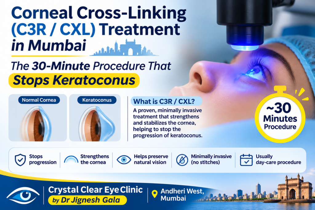
Corneal Cross-Linking (C3R/CXL) Treatment in Mumbai — The 30-Minute Procedure That Stops Keratoconus
Written & medically reviewed by Dr. Jignesh Gala, Vitreoretinal & Cataract Surgeon (FRCS Glasgow), Founder & Director — Crystal Clear Eye Clinic (Andheri West) and Topax Eye Hospital (Vile Parle East), Mumbai. Facts verified: 24 July 2026.
Thank you for reading this post, don't forget to subscribe!⚡ Quick answer: Corneal cross-linking (C3R / CXL) is the only proven treatment that stops keratoconus from progressing. Riboflavin (vitamin B2) drops are soaked into the cornea and activated with UV-A light for about 30 minutes, welding collagen fibres into a stiffer lattice so the cone stops bulging. It does not reverse the cone. For corneal cross-linking C3R CXL treatment in Mumbai with on-site tomography and progression-proven decisions, call 077188 85245.
- Singapore 2021 a year on the founding ophthalmology team of woodlands health
- Smartsurf with customeyes the most advanced glass removal surgery in mumbai and why it outperforms wavelight plus innoveyes
- Surgery packages
- Surgical videos by dr jignesh gala
- Types of intraocular lenses
- Types of refractive surgeries
- Types of retina surgeries
- Understanding and preventing digital eye strain
- Crystal Clear Eye Clinic — Home
- About Crystal Clear Eye Clinic, Andheri West
- Eye Health Blog — Crystal Clear Eye Clinic
- Contact & Directions — Andheri West
1. What Cross-Linking Is — Riboflavin + UV-A Stiffening the Cornea
Keratoconus is a biomechanical failure: the cornea’s collagen scaffolding is too weak, so normal eye pressure slowly pushes it outward into a cone. Corneal cross-linking (CXL) — known across India as C3R — attacks exactly that weakness. Riboflavin drops saturate the cornea; calibrated UV-A light then activates the vitamin, bonding adjacent collagen fibres into a stiffer lattice — cross-bracing for a sagging structure. The treated cornea resists further bulging; that is the entire goal.
Two honest truths first: CXL halts progression — it does not reverse the cone. Existing steepening largely stays; vision afterwards still comes from glasses, RGP or scleral lenses. And progression is provable, not opinion — serial corneal tomography (Kmax, ABCD grading) documents whether your cone is advancing. The decision here is made from numbers; stable corneas are honestly told they can wait.
Keratoconus is often discovered during a LASIK workup, when topography flags the cone. If that was your story: Contoura LASIK in Mumbai is strictly off-limits for keratoconic corneas — removing tissue from a weak cornea worsens it; adding bonds (CXL) stabilises it.
2. Epi-Off vs Epi-On vs Accelerated: The Protocol Question
Ask three Mumbai centres and you may hear three protocols. The honest 2026 evidence summary — the same comparison Dr. Gala walks patients through:
| Protocol | How It’s Done | Strength of Effect | Comfort & Recovery | Best Suited For |
|---|---|---|---|---|
| Conventional epi-off (Dresden) | Surface epithelium removed; riboflavin 30 min; UV-A 3 mW/cm² for 30 min | Gold standard — deepest, most proven stiffening; two decades of data | 2–4 days of real pain; bandage lens ~5 days; blurry first week | Clear documented progression, especially younger patients |
| Accelerated epi-off | Epithelium removed; higher UV-A power (9–30 mW/cm²) for 3–10 min — same energy, shorter time | Comparable halting in modern studies (2024 evidence, accelerated evidence) | Same healing as epi-off; chair time drops dramatically | Busy adults, bilateral cases, children |
| Epi-on (transepithelial) | Epithelium intact; enhanced riboflavin ± oxygen/iontophoresis penetrates through | Improving with newer high-fluence protocols, but on average less stiffening than epi-off | Minimal pain, fastest recovery, lowest haze/infection risk | Very thin corneas, patients who cannot afford downtime |
What this means in 2026: for a typical progressing adult, accelerated epi-off delivers Dresden-level stabilisation in a fraction of the chair time — Dr. Gala’s default. Epi-on suits thin corneas or minimal-downtime patients, with honest counselling about possible top-ups. Bilateral simultaneous accelerated protocols show solid 24-month results in children (Indian paediatric evidence, 2025).
One caution most clinics skip: RGP/hard lenses reshape the cornea and distort tomography — a proper CXL workup needs a 2-week lens washout before scanning.
3. The C3R Procedure Step by Step — 30–45 Minutes, Day Care
Patients ask C3R operation mein kya hota hai — the walkthrough:
- Pre-op scans: tomography and pachymetry confirm progression and safe corneal thickness (generally ≥400 microns).
- Numbed, not injected: anaesthetic drops numb the eye completely. No injection, no sedation; a small clip keeps the eyelids open.
- Riboflavin soaking (~10–30 min): in epi-off, the surface layer is gently removed first; riboflavin drops then saturate the cornea.
- UV-A activation (3–30 min by protocol): you stare at a soft blue-violet light while the UV-A source works. You feel nothing.
- Bandage lens + drops, then home: a protective soft lens goes on (epi-off); you leave within the hour.
The first week, honestly: days 1–3 after epi-off bring watering, burning and light sensitivity, managed with painkillers and the bandage lens. Vision blurs that week, clears over 1–4 weeks; desk work resumes in 4–7 days. Temporary dryness is common — the habits in our guide on preventing dry eyes help. Reviews at day 5, 1 month, then 6-monthly tomography prove the halt.
4. Who Needs Cross-Linking & When
The core principle: cross-linking treats progression, not the diagnosis. A keratoconus label alone is not an indication.
- Clear indication — documented progression: Kmax rising (~1 dioptre or more), thinning, or worsening vision on serial tomography. Once proven, every month of delay is cornea you do not get back.
- Young age = lower threshold: paediatric keratoconus progresses faster, so children are often cross-linked at diagnosis itself — reviews support early intervention (paediatric CXL review, paediatric meta-analysis), including eye-rubbing children with allergic eye disease (VKC + CXL evidence). Dr. Gala’s background as a paediatric ophthalmologist in Andheri, Mumbai means this urgency conversation happens in the first visit.
- Stable adult corneas can wait: unchanged tomography over a year? Monitor 6–12-monthly. No progression, no procedure.
- Too thin or too advanced: borderline corneas get modified protocols (epi-on, hypo-osmolar riboflavin); centrally scarred corneas may be beyond CXL’s benefit — that honesty is part of the consult.
What happens if you do nothing? Progressive keratoconus does not pause politely. It steepens until glasses fail, then lenses fail, and a minority — only about 10–15% of patients — end up needing a corneal transplant. Timely CXL is the single decision that most changes this disease’s lifetime trajectory.
5. CXL Cost in Mumbai — The 2026 Honest Band
Transparent, date-stamped pricing (verified 24 July 2026) — call 077188 85245 for an exact quote:
| Item | Mumbai 2026 Band | Notes |
|---|---|---|
| Corneal tomography / Pentacam scan | ₹1,200–2,500 (advanced tomography up to ₹5,000) | The mandatory baseline — and later the proof of the halt |
| CXL / C3R per eye | ₹25,000–45,000 typical (published Mumbai range ₹20,000–90,000) | Protocol (epi-off / accelerated / epi-on) and device generation drive the spread |
| Post-op medicines & bandage lens | Usually bundled or modest add-on | Confirm exactly what your quote includes |
| Follow-up tomography (6/12-month) | Scan rates as above | Non-negotiable — the proof progression has stopped |
Insurance & cashless: CXL is a surgical procedure covered under many Indian policies when progression is documented — unlike contact lenses, which are almost never covered. We supply the serial tomography and progression letters insurers ask for. On cashless: direct cashless is done through Topax Eye Care (Vile Parle East); third-party/TPA cashless is done through Crystal Clear Eye Clinic (Andheri West). And the economics: ₹25,000–45,000 once per eye now, versus a ₹75,000–1,50,000 corneal transplant later.
6. Why Patients Choose Dr. Jignesh Gala for Cross-Linking
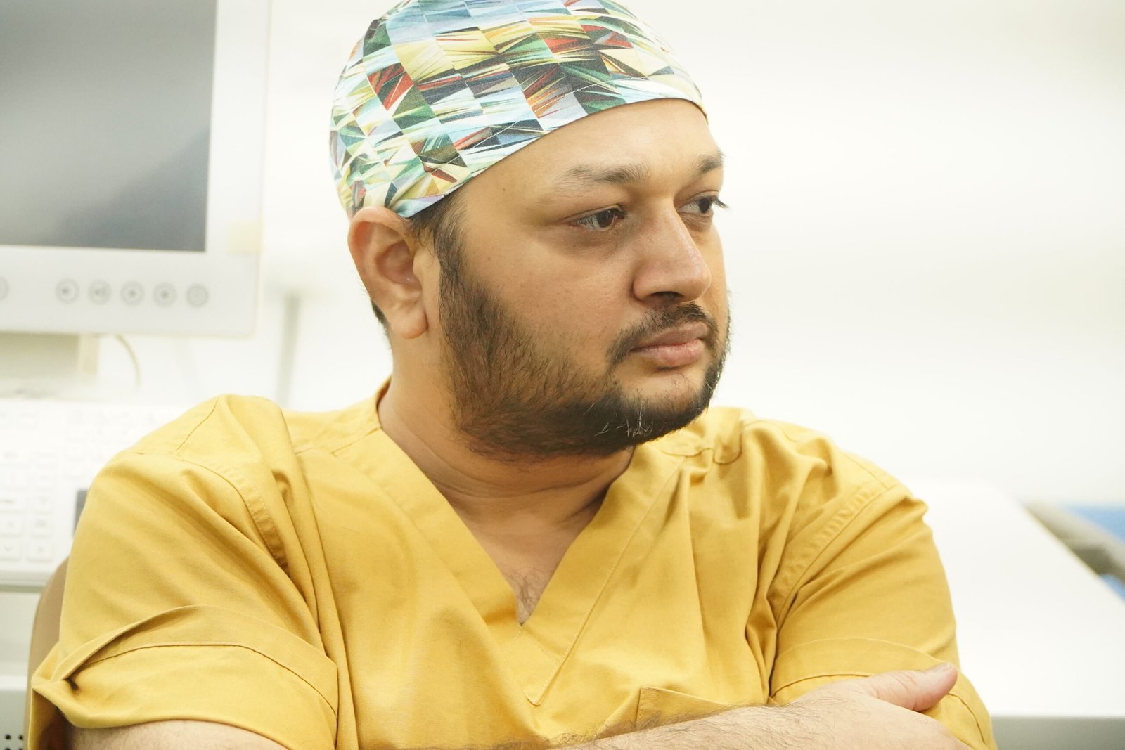
Dr. Jignesh Gala — FRCS (Glasgow), MRCS (Edinburgh), FICO; LVPEI fellowship; founding team at Woodlands Health, Singapore; former Assistant Professor at B.Y.L. Nair Hospital — owns both Crystal Clear Eye Clinic (Andheri West) and Topax Eye Hospital (Vile Parle East): the advice comes from the doctor accountable for the outcome.
✅ Progression-proven decisions: on-site tomography with serial Kmax/ABCD tracking — we cross-link when numbers demand it, and honestly tell stable corneas to wait.
✅ Protocol matched to your cornea: accelerated epi-off, conventional Dresden, or epi-on — chosen for your thickness, age and lifestyle.
✅ The complete keratoconus pathway under one roof: CXL at the centre, scleral-lens fitting coordinated with specialist fitters, Intacs evaluation and planning — and honest referral for true transplant-level cases.
✅ Fully aware of the latest research — unlike old practitioners who stopped reading journals years ago: he reads Cornea, JCRS, Journal of Refractive Surgery, AJO, BJO and IOVS — the journals this evidence lives in — and spent six years as a BMJ Case Reports peer reviewer. His training is documented openly: the FRCS Glasgow exam and the FICO 2016 exam.
✅ One accountable specialist, full scope: the same doctor Mumbai trusts as a top eye specialist in Mumbai — from glaucoma treatment and Eylea anti-VEGF injections to the HVF visual field test. Diabetic keratoconus patients get dual cornea-retina oversight from a diabetic retinopathy specialist in Andheri West; and if your eye ever signals danger — a curtain or shadow in your vision — you already have a surgeon who knows your eyes.
7. Locations — CXL in Andheri West & Vile Parle East
1) Crystal Clear Eye Clinic — Andheri West
📍 Address: Laram Centre CHS, A1-202, Swami Vivekanand Road, above Sunil Jewellers, near NADCO Shopping Centre, Railway Colony, Andheri West, Mumbai 400058
📞 Phone: 077188 85245 • 🕘 Open daily, closes 9 pm
💳 Third-party/TPA cashless is done through Crystal Clear Eye Clinic (Andheri West).
Minutes from Lokhandwala, Versova, Juhu, Oshiwara, DN Nagar and Jogeshwari; patients come from Bandra, Khar, Santacruz, Goregaon, Malad and Kandivali for corneal cross-linking C3R CXL treatment in Mumbai backed by on-site tomography.

2) Topax Eye Hospital — Vile Parle East
📍 Vile Parle East, Mumbai • 📞 077188 85245 • 🕘 Open daily, closes 9 pm
💳 Direct cashless is done through Topax Eye Care (Vile Parle East).
Convenient for Vile Parle, Santacruz East, Khar, Bandra East and the airport corridor — the complete CXL pathway under Dr. Gala.
8. हिंदी • मराठी • ગુજરાતી — Patient Summaries
🇮🇳 हिंदी सारांश — कॉर्नियल क्रॉस-लिंकिंग (C3R/CXL)
केराटोकोनस में कॉर्निया आगे उभरता है; C3R/CXL में राइबोफ्लेविन बूंदों और UV-A रोशनी से कॉर्निया मज़बूत होता है — यही इलाज प्रगति रोकता है (पुराना उभार वापस नहीं करता)। 30–45 मिनट की डे-केयर प्रक्रिया; मुंबई में ₹25,000–45,000 प्रति आँख (verified 24 July 2026)। कॉर्नियल क्रॉस-लिंकिंग C3R CXL ट्रीटमेंट मुंबई के लिए अपॉइंटमेंट: 077188 85245।
🚩 मराठी सारांश — कॉर्नियल क्रॉस-लिंकिंग (C3R/CXL)
केराटोकोनसमध्ये कॉर्निया पुढे फुगतो; C3R/CXL मध्ये रायबोफ्लेविन थेंब व UV-A किरणांनी कॉर्निया घट्ट होतो — हीच पद्धत प्रगती थांबवते (झालेला बदल उलटवत नाही). ३०–४५ मिनिटांची डे-केअर प्रक्रिया; मुंबईत ₹२५,०००–४५,००० प्रति डोळा (verified 24 July 2026). कॉर्नियल क्रॉस-लिंकिंग C3R CXL ट्रीटमेंट मुंबईसाठी भेट: 077188 85245.
🟡 ગુજરાતી સારાંશ — કોર્નિયલ ક્રોસ-લિંકિંગ (C3R/CXL)
કેરાટોકોનસમાં કોર્નિયા આગળ ફૂલે છે; C3R/CXLમાં રાઇબોફ્લેવિન ટીપાં અને UV-A પ્રકાશથી કોર્નિયા મજબૂત બને છે — આ જ ઈલાજ પ્રગતિ રોકે છે (જૂનો ફૂલાવો પાછો નથી લાવતો). ૩૦–૪૫ મિનિટની ડે-કેર પ્રક્રિયા; મુંબઈમાં ₹૨૫,૦૦૦–૪૫,૦૦૦ દરેક આંખ (verified 24 July 2026). કોર્નિયલ ક્રોસ-લિંકિંગ C3R CXL ટ્રીટમેન્ટ મુંબઈ માટે એપોઈન્ટમેન્ટ: 077188 85245.
9. ❓ 8 Frequently Asked Questions (English + Hinglish)
1. C3R / CXL kya hota hai?
C3R aur CXL ek hi ilaaj ke do naam hain. Keratoconus mein kamzor cornea ko riboflavin (vitamin B2) boondon se soak karke UV-A roshani se activate kiya jata hai — collagen fibres ke beech naye bonds bante hain aur cornea stiff ho jata hai. Result: progression rukti hai. Ye 30–45 minute ka day-care procedure hai.
2. Is corneal cross-linking painful?
The procedure itself is painless — anaesthetic drops numb the eye completely. The honest part: after epi-off CXL, days 1–3 bring burning, watering and light sensitivity while the surface heals; painkillers and a bandage lens control this. Epi-on is far more comfortable. Most resume routine work by day 4–7.
3. CXL cost in Mumbai kitna hai?
Mumbai mein typical cost ₹25,000–45,000 per eye hai (published range ₹20,000–90,000, verified 24 July 2026). Pehle corneal tomography (₹1,200–2,500) zaroori hoti hai. Many policies cover CXL as a surgical procedure when progression is documented. Exact quote: 077188 85245.
4. Does cross-linking improve vision?
Honestly: CXL’s job is to stop the disease getting worse, not to improve vision. Many eyes flatten slightly over the year — a bonus, not the promise. Sharp vision after CXL still usually comes from glasses, RGP or scleral lenses. The real win: the cornea you have today is the cornea you keep.
5. How long is recovery after cross-linking?
Epi-off: bandage lens ~5 days, discomfort days 1–3, blurry vision 1–4 weeks, desk work in 4–7 days. Epi-on: routine activity often in 1–2 days. We confirm the halt with tomography at 6 and 12 months.
6. Epi-off vs epi-on mein kya behtar hai?
Goal same, trade-off alag. Epi-off (including accelerated) deta hai sabse proven, sabse strong stiffening — lekin 2–4 din discomfort. Epi-on mein surface layer nahi hatati — dard kam, recovery fast — par average effect thoda kamzor aur kabhi-kabhi top-up lagta hai. Corneal thickness aur age ke hisaab se Dr. Gala honest recommendation dete hain.
7. Kya CXL se keratoconus ka progression ruk jata hai?
Haan — yehi iska maqsad hai, aur ye duniya ka sabse proven keratoconus treatment hai. Overwhelming majority of eyes mein progression rukti hai; ek chhota percentage mein top-up lag sakta hai. Isliye 6–12 mahine ki tomography follow-up compulsory hai — hum scan se prove karte hain, assume nahi.
8. Corneal cross-linking kahan hota hai Andheri West mein?
Crystal Clear Eye Clinic — Laram Centre CHS, A1-202, S.V. Road, above Sunil Jewellers, near NADCO Shopping Centre, Andheri West, Mumbai 400058 — on-site corneal tomography ke saath. Ya Topax Eye Hospital, Vile Parle East (direct cashless). Phone dono ka ek: 077188 85245. Open daily, closes 9 pm.
13. 📖 Related Reading — Complete Eye-Care Library by Dr. Jignesh Gala (78 guides)
Every guide below is written by the same surgeon who would operate on you:
From drjigneshgalaeye.com
- Drjigneshgalaeye.com
- A year at l v prasad eye institute how my vitreoretinal fellowship shaped the surgeon i am today
- About us
- Anti vegf injection cost in mumbai 2026 every drug every price explained honestly by a retina specialist
- B y l nair hospital 2019 2020 my sixteen months as assistant professor of ophthalmology
- Bakuchiol eye cream a gentle revolution in under eye care
- Best eye doctor for retina in mumbai dr jignesh gala retina vitreoretinal surgeon
- Best eye specialist in andheri mumbai
- Best retina specialist mumbai dr jignesh gala
- Best vision correction surgery after 40 what are your options
- Blogs by dr jignesh gala
- Cashless eye surgery in mumbai dr jignesh gala
- Diabetic retinopathy specialist in andheri west mumbai advanced screening injections laser vitrectomy by dr jignesh gala frcs
- Discover crystal clear vision with contoura lasik in mumbai
- Dr jignesh gala top eye specialist in mumbai
- Drjignesh gala premier pediatric ophthalmologistandheri mumbai
- Eylea injection in mumbai cost uses side effects the 2026 price revolution explained by a retina specialist
- Glaucoma treatment in andheri mumbai
- How to prevent dry eyes in winter
- Hvf test in andheri west mumbai visual field test cost procedure how to read your report by a specialist
- I see a curtain or shadow in my vision
- Inside my six years as a bmj case reports peer reviewer
- Inside the fico 2016 the only worldwide medical specialty examination
- Inside the frcs ophth glasgow how a 425 year old royal college tests your surgical knowledge
- Intravitreal injection in andheri west mumbai painless anti vegf eye injections by dr jignesh gala frcs
- Know your eye doctor
- Laser cataract surgery in andheri mumbai dr jignesh gala
- Laser cataract surgery
- Lasik eye surgery in andheri mumbai dr jignesh gala
- Lasik eye surgery in bandra mumbai dr jignesh gala
- Lasik eye surgery in juhu dr jignesh gala
- Lasik eye surgery in lokhandwala dr jignesh gala
- Lasik eye surgery in oshiwara dr jignesh gala
- Lasik in santacruz mumbai
- Lasik in versova mumbai dr jignesh gala
- Lasik surgery in andheri mumbai
- Most common types of refractive eye surgery which one is right for you
- Mrcs edinburgh how i joined a royal college founded in 1505
- Multifocal trifocal iol guide complete patient guide
- Oct eye test in andheri west mumbai cost procedure report why the specialist reading it matters
- People do ask google about this eye issue
- Refraction in real time how the royal college of ophthalmologists tested me in kuching 2017
- Refractive lens exchange rlethe complete guide to clear vision without glasses
- Retina laser treatment in andheri west mumbai prp barrage focal micropulse laser by dr jignesh gala frcs
- Retina surgery in mumbai your complete guide to saving your sight
- Retina treatment in andheri
- Retina treatment in mumbai advanced retinal care by dr jignesh gala
- Rle vs lasik which vision correction surgery is better
- Satisfied patient stories
- Singapore 2021 a year on the founding ophthalmology team of woodlands health
- Smartsurf with customeyes the most advanced glass removal surgery in mumbai and why it outperforms wavelight plus innoveyes
- Surgery packages
- Surgical videos by dr jignesh gala
- Types of intraocular lenses
- Types of refractive surgeries
- Types of retina surgeries
- Understanding and preventing digital eye strain
From crystalcleareye.in
- Crystal Clear Eye Clinic — Home
- About Crystal Clear Eye Clinic, Andheri West
- Eye Health Blog — Crystal Clear Eye Clinic
- Contact & Directions — Andheri West
- Glaucoma Treatment in Andheri, Mumbai
- LASIK Surgery in Andheri, Mumbai
- Premium IOL Selection Guide 2026
- Laser Cataract Surgery in Andheri, Mumbai
- Best Eye Specialist in Andheri, Mumbai
- Best Retina Specialist in Mumbai
- Best Vitrectomy Surgery in Mumbai — Expert Retinal Care
- Diabetic Eye Care in Mumbai
- Diabetic Retinopathy in Andheri
- Diabetic Macular Edema (DME) Treatment in Mumbai
- Macular Edema Treatment in Andheri West
- ARMD Treatment in Andheri West, Mumbai
- Retina Treatment in Andheri
- Vitreoretinal Treatment in Andheri
- Cashless Eye Surgery in Mumbai
- Sudden Floaters? Retina Check-up in Mumbai
- What Is an Intravitreal Eye Injection?
Your retina deserves a specialist. Today, not someday.
Cross-linking doesn’t improve vision — it saves it. The earlier the cornea is strengthened, the more vision there is to protect.
📍 Andheri West: Laram Centre CHS, A1-202, S.V. Road, above Sunil Jewellers, near NADCO Shopping Centre, Mumbai 400058
📍 Vile Parle East: Topax Eye Hospital • Open daily, closes 9 pm
Dr. Jignesh Gala, FRCS (Glasgow) — Vitreoretinal & Cataract Surgeon • drjigneshgalaeye.com • crystalcleareye.in
Medical disclaimer: This page is educational and does not replace a personal consultation. Prices are indicative, verified on 24 July 2026, and may change. Every eye is different — decisions are made only after examination by Dr. Jignesh Gala.
Research & Evidence Library — 105 Verified References
Verified live in July 2026 via official NCBI E-utilities — including research from Cornea, JCRS, Journal of Refractive Surgery, AJO, BJO, IOVS, Eye & Contact Lens, IJO, Acta Ophthalmologica, Graefe’s Archive and TVST. The same journals Dr. Gala reads. Click any title to check the science yourself.
KC: Corneal Cross-Linking: Long-Term Outcomes
- Long-term outcomes of corneal crosslinking. — Current opinion in ophthalmology, 2024
- Long term results of accelerated corneal collagen cross-linking in pediatric keratoconus. — European journal of ophthalmology, 2021
- Long-Term Outcomes After Corneal Cross-linking for Progressive Keratoconus and Corneal Ectasia: A 10-Year Follow-Up of the Pivotal Study. — Eye & contact lens, 2023
- Corneal Collagen Cross-Linking for Keratoconus in Pediatric Patients-Long-Term Results. — Cornea, 2017
- Short- and long-term safety and efficacy of corneal collagen cross-linking in progressive keratoconus: A systematic review and meta-analysis of randomized controlled trials. — Taiwan journal of ophthalmology, 2023
- Corneal Allogenic Intrastromal Ring Segments (CAIRS) Combined With Corneal Cross-linking for Keratoconus. — Journal of refractive surgery (Thorofare, N.J. : 1995), 2018
- Safety and Efficacy of Corneal Cross-Linking in Patients Affected by Keratoconus: Long-Term Results. — Medical sciences (Basel, Switzerland), 2023
- Long-term changes in crystalline lens transparency after accelerated transepithelial corneal cross-linking in patients with keratoconus. — European journal of ophthalmology, 2023
- Long-term Outcomes of Accelerated Corneal Cross-linking in the Treatment of Keratoconus: Comparison of Hypotonic Riboflavin Solution With Standard Riboflavin Solution. — Journal of refractive surgery (Thorofare, N.J. : 1995), 2020
- Long-term Evaluation of Corneal Biomechanical Properties After Corneal Cross-linking for Keratoconus: A 4-Year Longitudinal Study. — Journal of refractive surgery (Thorofare, N.J. : 1995), 2018
- Cutting-edge cross-linking biomaterials advancing ophthalmic therapeutics. — Progress in retinal and eye research, 2026
- Five-year corneal cross-linking outcomes: A Save Sight Keratoconus Registry Study. — Clinical & experimental ophthalmology, 2023
- Topography-guided photorefractive keratectomy combined with accelerated corneal collagen cross-linking versus cross-linking alone for progressive keratoconus: a long-term prospective cohort study. — Frontiers in medicine, 2024
- Long-term results of accelerated and conventional corneal cross-linking. — International ophthalmology, 2020
- Case for Epithelium-Off Corneal Cross-linking. — Cornea, 2022
KC: Accelerated Vs Conventional Cross-Linking (Epi-On)
- Keratoconus Treatment Algorithm. — Ophthalmology and therapy, 2017
- Expanding indications for corneal cross-linking. — Current opinion in ophthalmology, 2023
- Accelerated Epi-On Versus Standard Epi-Off Corneal Collagen Cross-Linking for Progressive Keratoconus in Pediatric Patients: Five Years of Follow-Up. — Cornea, 2020
- Accelerated Epi-On Versus Standard Epi-Off Corneal Collagen Cross-Linking for Progressive Keratoconus in Pediatric Patients. — Cornea, 2017
- A Transepithelial Corneal Cross-linking (CXL) Protocol Providing the Same Biomechanical Strengthening as Accelerated Epithelium-off CXL. — Journal of refractive surgery (Thorofare, N.J. : 1995), 2025
- A Review of Keratoconus Cross-Linking Treatment Methods. — Journal of clinical medicine, 2025
- Accelerated epithelium-on or accelerated epithelium-off corneal collagen cross-linking: Contralateral comparison study. — Taiwan journal of ophthalmology, 2020
- Progressive high-fluence epithelium-on accelerated corneal crosslinking: a novel corneal photodynamic therapy for early progressive keratoconus. — Frontiers in medicine, 2023
- Efficacy and safety of progressive fluence pulsed light epithelium-on accelerated corneal cross-linking for progressive keratoconus: 18-month prospective results. — International journal of ophthalmology, 2026
- Visual and Topographic Improvement with Epithelium-On, Oxygen-Supplemented, Customized Corneal Cross-Linking for Progressive Keratoconus. — Journal of clinical medicine, 2020
- Optimization of Oxygen Dynamics, UV-A Delivery, and Drug Formulation for Accelerated Epi-On Corneal Crosslinking. — Current eye research, 2020
- Epithelium-on Corneal Cross-linking Using a Modified Penetration Enhancer Without Supplemental Oxygen or Iontophoresis. — Journal of refractive surgery (Thorofare, N.J. : 1995), 2026
- Topo-Pachimetric Accelerated Epi-On Cross-Linking Compared to the Dresden Protocol Using Riboflavin with Vitamin E TPGS: Results of a 2-Year Randomized Study. — Journal of clinical medicine, 2021
- Management of Tomographically Suspicious Fellow Eyes of Young Patients With Unilateral Clinically Evident Keratoconus: Accelerated Epithelium-On Corneal Crosslinking Versus Observation. — Cornea, 2025
- Sterile corneal infiltrates after corneal collagen cross-linking: evaluation of risk factors. — Acta ophthalmologica, 2017
KC: Pediatric Keratoconus Cross-Linking
- Progression of Pediatric Keratoconus After Corneal Cross-Linking: A Systematic Review and Pooled Analysis. — Cornea, 2022
- Corneal Cross-Linking for Pediatric Keratoconus. — Diagnostics (Basel, Switzerland), 2024
- Accelerated Corneal Collagen Cross-Linking in Pediatric Keratoconus. — Journal of current ophthalmology, 2021
- Corneal collagen cross-linking in pediatric keratoconus with three protocols: a systematic review and meta-analysis. — Journal of AAPOS : the official publication of the American Association for Pediatric Ophthalmology and Strabismus, 2020
- Accelerated Corneal Cross-linking in Pediatric Patients with Progressive Keratoconus: 12-Month Follow-up Results. — Nigerian journal of clinical practice, 2024
- Investigation of Epithelial Remodeling After Accelerated Epi-Off Cross-linking in Pediatric Keratoconus. — Eye & contact lens, 2025
- Corneal Cross-Linking as Treatment in Pediatric Keratoconus: Comparison of Two Protocols. — Journal of ophthalmology, 2021
- Corneal Collagen Cross-Linking for Progressive Keratoconus in Pediatric Patients: Up to 14 Years of Follow-up. — American journal of ophthalmology, 2023
- Astigmatic Vectorial Analysis in Pediatric Keratoconus After Unilateral Cross-Linking Treatment. — Cornea, 2022
- Corneal Cross-Linking for Pediatric Keratcoconus Review. — Cornea, 2018
- Outcomes of accelerated corneal cross-linking for pediatric and adult keratoconus: a comparative study. — International ophthalmology, 2024
- Corneal Cross-Linking for Paediatric Keratoconus: A Systematic Review and Meta-Analysis. — Journal of clinical medicine, 2021
- Comparison of Two Different Accelerated Corneal Cross-linking Procedure Outcomes in Patients with Keratoconus. — Balkan medical journal, 2020
- Bilateral simultaneous accelerated corneal collagen cross-linking in pediatric keratoconus: 24-month follow-up results. — Indian journal of ophthalmology, 2025
- Safety and Efficacy of Corneal Cross-linking in Pediatric Patients with Keratoconus and Vernal Keratoconjunctivitis. — Middle East African journal of ophthalmology, 2019
KC: Scleral Lenses: Visual Rehabilitation
- Keratoconus Diagnosis and Treatment: Recent Advances and Future Directions. — Clinical ophthalmology (Auckland, N.Z.), 2023
- Visual rehabilitation with mini scleral contact lenses in scarred corneas. — Journal francais d’ophtalmologie, 2024
- Efficacy of mini-scleral lenses in visual rehabilitation for corneal disorders: a prospective study. — BMC ophthalmology, 2025
- Visual rehabilitation using SPOT(®) scleral lenses in patients with keratoconus. — European journal of ophthalmology, 2026
- Visual rehabilitation using mini-scleral contact lenses after penetrating keratoplasty. — Arquivos brasileiros de oftalmologia, 2017
- Refractive approaches to visual rehabilitation in patients with keratoconus. — Current opinion in ophthalmology, 2020
- Visual Rehabilitation of Bilateral Keratoconus With Customized Scleral Lenses: A Case Report. — Cureus, 2026
- Safety and Efficacy of Scleral Lenses for Keratoconus. — Optometry and vision science : official publication of the American Academy of Optometry, 2020
- Contact lenses for visual rehabilitation in post-keratoplasty eyes: A review. — Indian journal of ophthalmology, 2025
- Scleral contact lenses for visual rehabilitation after penetrating keratoplasty: long term outcomes. — Contact lens & anterior eye : the journal of the British Contact Lens Association, 2014
- Mini-Scleral Lens: An Effective Mode of Visual Rehabilitation in Eyes With Resolved Hydrops. — Eye & contact lens, 2024
- Influence of Scleral Contact Lenses on Optical Coherence Tomography Parameters in Keratoconus Patients. — Diagnostics (Basel, Switzerland), 2025
- Impact of Scleral Lenses on Visual Acuity and Ocular Aberrations in Corneal Ectasia: A Comprehensive Review. — Journal of personalized medicine, 2024
- Irregular Corneas: Improve Visual Function With Scleral Contact Lenses. — Eye & contact lens, 2018
- Visual performance of scleral lenses and their impact on quality of life in patients with irregular corneas. — Arquivos brasileiros de oftalmologia, 2018
KC: Intracorneal Ring Segments (Intacs)
- Intracorneal Ring Segments in Keratoconus: A Narrative Literature Review. — Clinical ophthalmology (Auckland, N.Z.), 2025
- Intracorneal ring segment implantation in advanced Keratoconus. — European journal of ophthalmology, 2023
- Clinical Outcomes and Complications After INTACS Implantation in Keratoconus: A Systematic Review. — Journal of clinical medicine, 2026
- Refractive and aberrometric outcomes of intracorneal ring segments for keratoconus: mechanical versus femtosecond-assisted procedures. — Ophthalmology, 2009
- [Intacsintracorneal ring segments in keratoconus]. — Der Ophthalmologe : Zeitschrift der Deutschen Ophthalmologischen Gesellschaft, 2013
- Outcomes of Single Segment Implantation of Conventional Intacs versus Intacs SK for Keratoconus. — Journal of ophthalmic & vision research, 2014
- Implantation of intracorneal ring segments in pediatric patients: long-term follow-up. — International medical case reports journal, 2018
- Intracorneal ring segments for keratoconus correction: long-term follow-up. — Journal of cataract and refractive surgery, 2006
- Intracorneal ring segments in ectatic corneal disease – a review. — Clinical & experimental ophthalmology, 2010
- Intracorneal Ring Segments Followed by Simultaneous Topography-Guided Removal of Epithelium and Stroma With Accelerated Collagen Cross-Linking For Keratoconus (I-TRESK/CXL). — Asia-Pacific journal of ophthalmology (Philadelphia, Pa.), 2020
- Intacs in advanced keratoconus. — Cornea, 2008
- Intracorneal ring segment implantation for the management of keratoconus: safety and efficacy. — Journal of cataract and refractive surgery, 2007
- Corneal crosslinking and intracorneal ring segments for keratoconus: A randomized study of concurrent versus sequential surgery. — Journal of cataract and refractive surgery, 2019
- Long-term follow up of intracorneal ring segment implantation in 932 keratoconus eyes. — Journal francais d’ophtalmologie, 2020
- Modified intracorneal ring segment implantations (INTACS) for the management of moderate to advanced keratoconus: efficacy and complications. — Cornea, 2006
KC: Genetics, Pathophysiology & Biomechanics
- Advances in keratoconus animal models: From genetics to biomechanics. — Experimental eye research, 2025
- A review of keratoconus: Diagnosis, pathophysiology, and genetics. — Survey of ophthalmology, 2017
- [KERATOCONUS: DIAGNOSIS AND INNOVATIONS]. — Harefuah, 2025
- The pathogenesis of keratoconus. — Eye (London, England), 2014
- Nutritional and Metabolic Imbalance in Keratoconus. — Nutrients, 2022
- Update on the Physiopathology of Keratoconus. — Medical sciences (Basel, Switzerland), 2026
- The Proteins of Keratoconus: a Literature Review Exploring Their Contribution to the Pathophysiology of the Disease. — Advances in therapy, 2019
- Experimental models for keratoconus: Insights and challenges. — Experimental eye research, 2024
- Tear Proteomic Analysis From Offspring of Keratoconus Patients: New Insights Into Corneal Biomechanical Weakness and Disease Risk Stages. — Investigative ophthalmology & visual science, 2025
- Biomechanical and inflammatory pathways underlying the genetic architecture of keratoconus: A genomic SEM study. — Experimental eye research, 2026
- The importance of corneal biomechanics in assessing first degree family members of keratoconus patients. — Romanian journal of ophthalmology, 2018
- Keratoconus Prevalence in Families: A French Study. — Cornea, 2020
- Fourteen years follow-up of a stable unilateral Keratoconus: unique case report of clinical, tomographical and biomechanical stability. — BMC ophthalmology, 2022
- Genetic Variants Associated With Corneal Biomechanical Properties and Potentially Conferring Susceptibility to Keratoconus in a Genome-Wide Association Study. — JAMA ophthalmology, 2019
- Update on the genetics of keratoconus. — Experimental eye research, 2021
KC: Corneal Allogenic Intrastromal Ring Segments (Cairs)
- Customized corneal allogenic intrastromal ring segments (CAIRS) for keratoconus with decentered asymmetric cone. — Indian journal of ophthalmology, 2023
- Corneal Allogenic Intrastromal Ring Segments (CAIRS) Versus Synthetic Segments: A Single Segment Comparative Analysis Using Propensity Score Matching. — Journal of refractive surgery (Thorofare, N.J. : 1995), 2024
- Corneal Allogenic Intrastromal Ring Segments: A Literature Review. — Journal of clinical medicine, 2025
- Corneal Allogeneic Intrastromal Ring Segments (CAIRS): The New Kid on the Block. — Klinische Monatsblatter fur Augenheilkunde, 2025
- Visual and Topographic Outcomes After Corneal Allogeneic Intrastromal Ring Segments for Keratoconus: A Systematic Review and Meta-Analysis. — American journal of ophthalmology, 2025
- Customized Corneal Allogeneic Intrastromal Ring Segments (CAIRS) With Corneal Cross-linking for Post-LASIK Ectasia. — Journal of refractive surgery (Thorofare, N.J. : 1995), 2025
- Extracorporeal Optimization of Corneal Allogenic Intrastromal Ring Segments (ECO-CAIRS) Using Ultra-high-Fluence Corneal Cross-linking. — Journal of refractive surgery (Thorofare, N.J. : 1995), 2025
- Corneal Allogeneic Intrastromal Ring Segments for keratoconus – recent evidence and the move to customized and custom shaped CAIRS. — Current opinion in ophthalmology, 2026
- Femtosecond Laser Created Corneal Allogenic Intrastromal Ring Segments for Keratoconus. — Clinical & experimental ophthalmology, 2026
- Corneal Allogeneic Intrastromal Ring Segments (CAIRS): Indications, Mechanism of Action and Outcomes. — Klinische Monatsblatter fur Augenheilkunde, 2026
- Corneal allogenic intrastromal ring segments improve visual and topographic outcomes in advanced keratoconus : Short Title: CAIRS in Advanced Keratoconus. — Scientific reports, 2026
- Effect of Corneal Allogenic Intrastromal Ring Segment (CAIRS) Implantation Surgery in Patients With Keratoconus According to Prior Corneal Cross-linking Status. — Journal of refractive surgery (Thorofare, N.J. : 1995), 2024
- Intrastromal corneal ring segments for keratoconus: a comprehensive review of different types. — Therapeutic advances in ophthalmology, 2025
- Postoperative Adjustment of Implanted Corneal Allogeneic Intrastromal Ring Segments (CAIRS) to Improve Visual and Topographic Outcomes. — Journal of refractive surgery (Thorofare, N.J. : 1995), 2025
- Use of corneal allogenic intrastromal ring segments (CAIRS) in high astigmatism, a case report. — American journal of ophthalmology case reports, 2026