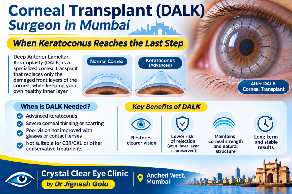
Corneal Transplant (DALK) Surgeon in Mumbai — When Keratoconus Reaches the Last Step
Written & medically reviewed by Dr. Jignesh Gala, Vitreoretinal & Cataract Surgeon (FRCS Glasgow), Founder & Director — Crystal Clear Eye Clinic (Andheri West) and Topax Eye Hospital (Vile Parle East), Mumbai. Facts verified: 24 July 2026.
Thank you for reading this post, don't forget to subscribe!⚡ Quick answer: Searching for a corneal transplant (DALK) surgeon in Mumbai because keratoconus has advanced? Know this first: only about 10–15% of keratoconus patients ever need a transplant. The honest sequence is cross-linking to stop progression, scleral lenses or Intacs to restore vision — and transplant (DALK or PK) only when those genuinely fail. At Crystal Clear Eye Clinic, Andheri West, Dr. Jignesh Gala (FRCS Glasgow) gives you the honest evaluation: serial tomography, every alternative exhausted first, and for true transplant cases, planned referral to a corneal transplant specialist with full co-management before and after surgery. DALK/transplant band in Mumbai: ₹75,000–1,50,000 (verified 24 July 2026). Call 077188 85245.
- I see a curtain or shadow in my vision
- Inside my six years as a bmj case reports peer reviewer
- Inside the fico 2016 the only worldwide medical specialty examination
- Inside the frcs ophth glasgow how a 425 year old royal college tests your surgical knowledge
- Intravitreal injection in andheri west mumbai painless anti vegf eye injections by dr jignesh gala frcs
- Know your eye doctor
- Laser cataract surgery in andheri mumbai dr jignesh gala
- Laser cataract surgery
- Lasik eye surgery in andheri mumbai dr jignesh gala
- Lasik eye surgery in bandra mumbai dr jignesh gala
- Lasik eye surgery in juhu dr jignesh gala
- Lasik eye surgery in lokhandwala dr jignesh gala
1. When Keratoconus Needs a Transplant (the Honest 10–15%)
Keratoconus thins and steepens the cornea into a cone. Cross-linking stops the worsening; lenses work around the shape; a transplant replaces the cornea itself — which is why it stays the last step, reserved for honest indications:
- Corneal scarring after acute hydrops — a tear in the inner layer floods the cornea; the healed scar can block central vision permanently.
- Extreme thinning — too thin to cross-link safely, or too steep for any lens to sit.
- Contact-lens failure — genuine intolerance or a cone too advanced for sclerals, after a proper specialist fitting trial (not one rushed attempt).
- Vision no optical device can restore — glasses fail, lenses fail, and the cornea itself is the wall.
Everything short of that list has a less invasive answer. Scleral lenses vault even severely irregular corneas and improve acuity and aberrations (2024 comprehensive review, irregular-cornea evidence), with real quality-of-life gains (2018 QoL study). Intacs help selected advanced corneas (advanced keratoconus ICRS 2023, 2025 narrative review). Reaching for transplant before exhausting these is not thoroughness — it is haste.
2. DALK vs PK: The Honest Comparison
Two operations dominate keratoconus transplantation. The difference is which layers of the cornea are replaced — and that changes rejection risk, recovery and long-term strength:
| DALK (Deep Anterior Lamellar Keratoplasty) | PK (Penetrating Keratoplasty) | |
|---|---|---|
| What is replaced | Front ~90% of cornea (epithelium + stroma); your own healthy endothelium is kept | Full-thickness cornea — all layers including endothelium |
| Rejection profile | Much lower — endothelial rejection (the dangerous kind) largely eliminated | Higher — endothelial rejection possible lifelong; steroid drops often for years |
| Recovery | Rehabilitation over months; sutures adjusted/removed over 12–18 months | Similar timeline; refraction unstable longer |
| Sutures | Present — follow-up for adjustment/removal mandatory | Present; removal schedule similar |
| Eye strength after healing | Stronger — your own inner layer preserves structural integrity | Full-thickness wound margin remains a lifelong weak point; trauma risk higher |
| Best suited when | Stromal disease with a healthy endothelium — most keratoconus cases | Endothelium also damaged (e.g., after severe hydrops), deep scars crossing layers |
The honest summary: for typical keratoconus, DALK is preferred when achievable because it keeps your endothelium — the cells that keep the cornea clear, whose loss drives PK graft failure. PK answers when disease crosses all layers. Either way, grafts often still need scleral lenses afterward — transplant resets anatomy, not optics.
3. The Transplant Pathway — What Patients Must Commit To
- Evaluation (weeks): serial tomography to prove the cornea — not the lens fit — is the limiting factor; endothelial assessment; documentation of alternatives tried.
- Listing & tissue: registration with the transplant centre and eye bank; donor tissue takes days to weeks.
- Surgery (day): a few hours under anaesthesia at a transplant-equipped hospital; day-care or one-night stay.
- The long follow-up (12–18 months): reviews at days, weeks, then monthly; suture adjustments and staged removal; rejection vigilance; finally optical rehabilitation — often scleral lenses over the graft. Sclerals influence measurable OCT parameters in keratoconus (2025 Diagnostics study), so fitting on a graft is specialist work.
What you must commit to before saying yes: 12–18 months of scheduled follow-ups, years of anti-rejection drops (especially after PK), protection from eye trauma, and accepting that final vision often comes from a lens over the graft. A transplant is an 18-month project with a lifelong maintenance chapter — not a one-day fix.
4. Our Role: Honest Evaluation, Exhausting Alternatives First, Co-Managed Referral
Let us be precise about what we do — and do not claim:
- We evaluate honestly. Serial tomography (Kmax/ABCD), pachymetry, refraction history and lens trials answer the only question that matters: is transplant truly the remaining option? Many “you need a transplant” opinions dissolve once a proper scleral fitting is actually attempted.
- We exhaust alternatives first. Cross-linking (C3R/CXL) at our centre, specialist scleral-lens fitting, Intacs evaluation & planning — in that order, with the evidence above behind each.
- For true transplant cases, we refer — planned, not abandoned. Dr. Gala does not perform corneal transplant surgery himself; DALK/PK belongs with a dedicated corneal transplant surgeon at an eye-bank-backed hospital. What we do: prepare your complete record set, refer you to the right transplant specialist, and co-manage — pre-op optimization plus the long post-op follow-up (suture visits, surface care, scleral-lens rehabilitation over the graft) back at our Andheri West centre, minutes from home.
This model serves you twice: the transplant decision is made by someone with no incentive toward it, and the 18-month follow-up happens where you can actually attend it.
5. Corneal Transplant Cost in Mumbai (2026 Bands)
| Item | Mumbai 2026 Band | Notes |
|---|---|---|
| DALK / corneal transplant (per eye) | ₹75,000–1,50,000 | Surgeon, hospital, tissue charges; varies with DALK vs PK and hospital tier |
| Pre-transplant alternatives | CXL ₹25,000–45,000/eye (published range ₹20,000–90,000); scleral lenses ₹15,000–50,000/pair + fitting ₹500–3,000; Intacs ₹60,000–1,20,000/eye | Exhaust first — clinically and financially |
| Diagnostics | Corneal tomography ₹1,200–2,500 (advanced up to ₹5,000) | Serial scans needed before and after |
All bands verified 24 July 2026 — call 077188 85245 for an exact quote for your case.
Insurance reality: corneal transplant is medically necessary surgery and is covered under most standard policies; CXL is covered in many. Contact lenses are usually not covered. Cashless mechanics at our centres: direct cashless is done through Topax Eye Care (Vile Parle East); third-party/TPA cashless is done through Crystal Clear Eye Clinic (Andheri West). For transplant claims we document the medical-necessity trail — serial scans, lens-trial records — that claim desks ask for.
6. Why Patients Trust Dr. Jignesh Gala With the Transplant Decision

Dr. Jignesh Gala, FRCS (Glasgow), MRCS (Edinburgh), FICO — Founder & Director of Crystal Clear Eye Clinic (Andheri West) and Topax Eye Hospital (Vile Parle East); LVPEI fellowship; Woodlands Health Singapore founding team; ex-Assistant Professor, B.Y.L. Nair Hospital; 6 years a BMJ Case Reports peer reviewer. Patients know him as a leading eye specialist in Andheri and retina specialist in Mumbai — a surgeon who operates daily (vitrectomy, laser cataract) and therefore respects exactly when not to.
✅ Zero incentive toward transplant: because graft surgery goes to a transplant specialist, our evaluation has one loyalty — your cornea.
✅ Complete pre-transplant arsenal on-site: tomography with serial progression records, CXL at the centre, scleral-lens coordination, Intacs planning.
✅ Fully aware of the latest research — unlike old practitioners who stopped reading journals years ago: Dr. Gala reads Cornea, JCRS, Journal of Refractive Surgery, AJO, BJO and IOVS.
✅ Co-management that actually happens: structured handover to the transplant surgeon, then your 12–18 months of follow-up and lens rehabilitation back near home — the part of transplant care patients usually lose. Our wider surgical ecosystem — glaucoma, LASIK, premium IOL planning — keeps later eye decisions under one roof.
7. Locations — Andheri West & Vile Parle East
1) Crystal Clear Eye Clinic — Andheri West
📍 Address: Laram Centre CHS, A1-202, Swami Vivekanand Road, above Sunil Jewellers, near NADCO Shopping Centre, Railway Colony, Andheri West, Mumbai 400058 • Open daily, closes 9 pm.
📞 Phone: 077188 85245 • 💬 WhatsApp Us
💳 Direct cashless is done through Topax Eye Care (Vile Parle East); third-party/TPA cashless is done through Crystal Clear Eye Clinic (Andheri West).
Minutes from Lokhandwala, Versova, Juhu, Oshiwara, DN Nagar and Jogeshwari; 20–35 minutes from Bandra, Khar, Santacruz, Goregaon, Malad and Kandivali. Post-graft follow-up is exactly where a near-home centre pays off. We also manage the conditions that travel with transplant patients: diabetic eye care, diabetic retinopathy, diabetic macular edema, macular edema and ARMD treatment in Andheri West.

2) Topax Eye Hospital — Vile Parle East
📍 Vile Parle East, Mumbai • 📞 077188 85245
💳 Direct cashless is done through Topax Eye Care. Hospital-grade setting convenient for Santacruz East, Khar East, Kalina and the airport corridor.
8. हिंदी • मराठी • ગુજराती — Patient Summaries
🇮🇳 हिंदी सारांश
Corneal transplant DALK surgeon Mumbai खोज रहे हैं? पहले यह जानें — सिर्फ 10–15% keratoconus patients को transplant चाहिए। डॉ. जिग्नेश गाला ईमानदार evaluation करते हैं: पहले CXL, scleral lenses और Intacs; सच्चे cases में specialist को planned referral और co-management। खर्च ₹75,000–1,50,000 (verified 24 July 2026)। अपॉइंटमेंट: 077188 85245।
🚩 मराठी सारांश
Corneal transplant DALK surgeon Mumbai शोधत असाल? फक्त १०–१५% किराटोकोनस रुग्णांना ग्राफ्ट लागते. डॉ. जिग्नेश गाला प्रामाणिक मूल्यमापन करतात — आधी CXL, स्क्लेरल लेन्स व Intacs; खरी गरज असल्यास ट्रान्सप्लांट तज्ज्ञाकडे नियोजित संदर्भ व सह-व्यवस्थापन. खर्च ₹७५,०००–१,५०,०००. भेटीसाठी: 077188 85245.
🟡 ગુજરાતી સારાંશ
Corneal transplant DALK surgeon Mumbai શોધો છો? ફક્ત ૧૦–૧૫% કેરાટોકોનસ દર્દીઓને ટ્રાન્સપ્લાન્ટ જોઈએ. ડૉ. જિગ્નેશ ગાલા પ્રમાણિક મૂલ્યાંકન કરે છે — પહેલા CXL, સ્ક્લેરલ લેન્સ અને Intacs; ખરી જરૂર હોય તો નિષ્ણાત પાસે આયોજિત રેફરલ અને સહ-સંચાલન. ખર્ચ ₹૭૫,૦૦૦–૧,૫૦,૦૦૦. એપોઈન્ટમેન્ટ: 077188 85245.
9. ❓ Frequently Asked Questions
1. DALK surgery kya hoti hai?
DALK (Deep Anterior Lamellar Keratoplasty) mein cornea ke front layers donor tissue se replace hote hain, par aapka healthy inner layer (endothelium) bacha rehta hai. Isliye rejection ka khatra PK se bahut kam — aur ye keratoconus ka preferred technique hai jab endothelium theek ho.
2. Kitne percent keratoconus patients ko transplant chahiye?
Sirf lagbhag 10–15%. Baaki 85–90% cross-linking (progression rokne ke liye), scleral lenses (vision ke liye) aur kabhi-kabhi Intacs se manage ho jaate hain. Agar kisi ne pehli visit mein hi transplant bola, toh serial scans aur proper lens trial maangiye — phir decide kijiye.
3. Corneal transplant cost in Mumbai kitna hai?
DALK/corneal transplant: ₹75,000–1,50,000 per eye (verified 24 July 2026) — surgeon, hospital aur tissue charges ke hisaab se. Alternatives ka kharcha: CXL ₹25,000–45,000/eye, scleral lenses ₹15,000–50,000/pair, Intacs ₹60,000–1,20,000/eye. Transplant aur CXL kai policies mein covered hain; lenses usually nahi. Exact quote: 077188 85245.
4. DALK vs PK mein kya farak hai?
DALK sirf front layers replace karta hai (aapki endothelium bachi rehti hai) — rejection risk kam, aankh stronger. PK poori thickness replace karta hai — zaroori jab inner layer bhi kharab ho, par lifelong rejection risk zyada. Dono mein sutures aur 12–18 mahine follow-up common hai. Full table Section 2 mein hai.
5. How long is recovery after DALK?
Think in months, not days: usable vision often returns within weeks, but sutures are adjusted and removed over 12–18 months, and best vision typically arrives only after suture removal — often with a scleral lens over the graft. Commit to the full follow-up schedule before choosing surgery.
6. Kya transplant ke baad vision normal ho jati hai?
Honestly — “normal” rare hai. Graft cornea ko clear banata hai, par perfectly regular nahi; aksar best vision scleral lens ke saath aati hai. Expectation: cloudy cornea se clear cornea + lens-assisted good functional vision. Jo “bilkul normal” promise kare, usse written mein poochhiye.
7. Corneal transplant surgeon Mumbai mein kaun ache hain?
Seedha jawab: Dr. Gala khud corneal transplant surgery nahi karte — graft surgery dedicated corneal transplant surgeons aur eye-bank-backed hospitals ka kaam hai. Humari bhoomika: honest evaluation, saare alternatives exhaust karna, phir sahi transplant specialist ko planned referral — aur 12–18 mahine ka follow-up humare Andheri West centre par. Ye model aapko turant surgery ke incentive se bachata hai.
8. Do you evaluate transplant-necessity honestly?
Yes — that is the design of our service. Serial tomography proves whether the cornea is truly end-stage; specialist scleral-lens fitting is genuinely attempted; Intacs is considered where suitable. Only when the data says “transplant” do we say it — then we refer and co-manage. Bring old scans and any surgery advice note: 077188 85245.
13. 📖 Related Reading — Complete Eye-Care Library by Dr. Jignesh Gala (78 guides)
Every guide below is written by the same surgeon who would operate on you:
From drjigneshgalaeye.com
- Drjigneshgalaeye.com
- A year at l v prasad eye institute how my vitreoretinal fellowship shaped the surgeon i am today
- About us
- Anti vegf injection cost in mumbai 2026 every drug every price explained honestly by a retina specialist
- B y l nair hospital 2019 2020 my sixteen months as assistant professor of ophthalmology
- Bakuchiol eye cream a gentle revolution in under eye care
- Best eye doctor for retina in mumbai dr jignesh gala retina vitreoretinal surgeon
- Best eye specialist in andheri mumbai
- Best retina specialist mumbai dr jignesh gala
- Best vision correction surgery after 40 what are your options
- Blogs by dr jignesh gala
- Cashless eye surgery in mumbai dr jignesh gala
- Diabetic retinopathy specialist in andheri west mumbai advanced screening injections laser vitrectomy by dr jignesh gala frcs
- Discover crystal clear vision with contoura lasik in mumbai
- Dr jignesh gala top eye specialist in mumbai
- Drjignesh gala premier pediatric ophthalmologistandheri mumbai
- Eylea injection in mumbai cost uses side effects the 2026 price revolution explained by a retina specialist
- Glaucoma treatment in andheri mumbai
- How to prevent dry eyes in winter
- Hvf test in andheri west mumbai visual field test cost procedure how to read your report by a specialist
- I see a curtain or shadow in my vision
- Inside my six years as a bmj case reports peer reviewer
- Inside the fico 2016 the only worldwide medical specialty examination
- Inside the frcs ophth glasgow how a 425 year old royal college tests your surgical knowledge
- Intravitreal injection in andheri west mumbai painless anti vegf eye injections by dr jignesh gala frcs
- Know your eye doctor
- Laser cataract surgery in andheri mumbai dr jignesh gala
- Laser cataract surgery
- Lasik eye surgery in andheri mumbai dr jignesh gala
- Lasik eye surgery in bandra mumbai dr jignesh gala
- Lasik eye surgery in juhu dr jignesh gala
- Lasik eye surgery in lokhandwala dr jignesh gala
- Lasik eye surgery in oshiwara dr jignesh gala
- Lasik in santacruz mumbai
- Lasik in versova mumbai dr jignesh gala
- Lasik surgery in andheri mumbai
- Most common types of refractive eye surgery which one is right for you
- Mrcs edinburgh how i joined a royal college founded in 1505
- Multifocal trifocal iol guide complete patient guide
- Oct eye test in andheri west mumbai cost procedure report why the specialist reading it matters
- People do ask google about this eye issue
- Refraction in real time how the royal college of ophthalmologists tested me in kuching 2017
- Refractive lens exchange rlethe complete guide to clear vision without glasses
- Retina laser treatment in andheri west mumbai prp barrage focal micropulse laser by dr jignesh gala frcs
- Retina surgery in mumbai your complete guide to saving your sight
- Retina treatment in andheri
- Retina treatment in mumbai advanced retinal care by dr jignesh gala
- Rle vs lasik which vision correction surgery is better
- Satisfied patient stories
- Singapore 2021 a year on the founding ophthalmology team of woodlands health
- Smartsurf with customeyes the most advanced glass removal surgery in mumbai and why it outperforms wavelight plus innoveyes
- Surgery packages
- Surgical videos by dr jignesh gala
- Types of intraocular lenses
- Types of refractive surgeries
- Types of retina surgeries
- Understanding and preventing digital eye strain
From crystalcleareye.in
- Crystal Clear Eye Clinic — Home
- About Crystal Clear Eye Clinic, Andheri West
- Eye Health Blog — Crystal Clear Eye Clinic
- Contact & Directions — Andheri West
- Glaucoma Treatment in Andheri, Mumbai
- LASIK Surgery in Andheri, Mumbai
- Premium IOL Selection Guide 2026
- Laser Cataract Surgery in Andheri, Mumbai
- Best Eye Specialist in Andheri, Mumbai
- Best Retina Specialist in Mumbai
- Best Vitrectomy Surgery in Mumbai — Expert Retinal Care
- Diabetic Eye Care in Mumbai
- Diabetic Retinopathy in Andheri
- Diabetic Macular Edema (DME) Treatment in Mumbai
- Macular Edema Treatment in Andheri West
- ARMD Treatment in Andheri West, Mumbai
- Retina Treatment in Andheri
- Vitreoretinal Treatment in Andheri
- Cashless Eye Surgery in Mumbai
- Sudden Floaters? Retina Check-up in Mumbai
- What Is an Intravitreal Eye Injection?
Your retina deserves a specialist. Today, not someday.
Only about 10–15% of keratoconus ever needs a transplant — but if you do, you need the truth, not the sales pitch.
📍 Andheri West: Laram Centre CHS, A1-202, S.V. Road, above Sunil Jewellers, near NADCO Shopping Centre, Mumbai 400058
📍 Vile Parle East: Topax Eye Hospital • Open daily, closes 9 pm
Dr. Jignesh Gala, FRCS (Glasgow) — Vitreoretinal & Cataract Surgeon • drjigneshgalaeye.com • crystalcleareye.in
Medical disclaimer: This page is educational and does not replace a personal consultation. Prices are indicative, verified on 24 July 2026, and may change. Every eye is different — decisions are made only after examination by Dr. Jignesh Gala.
Research & Evidence Library — 115 Verified References
Verified live in July 2026 via official NCBI E-utilities — including research from Cornea, JCRS, Journal of Refractive Surgery, AJO, BJO, IOVS, Eye & Contact Lens, IJO, Acta Ophthalmologica, Graefe’s Archive and TVST. The same journals Dr. Gala reads. Click any title to check the science yourself.
KC: Dalk Vs Penetrating Keratoplasty
- Penetrating Keratoplasty versus Deep Anterior Lamellar Keratoplasty for Keratoconus: A Systematic Review and Meta-analysis. — Journal of ophthalmic & vision research, 2022
- Penetrating Keratoplasty versus Deep Anterior Lamellar Keratoplasty for Keratoconus: A Systematic Review and Meta-Analysis of 27,018 Eyes. — Seminars in ophthalmology, 2025
- Deep anterior lamellar keratoplasty versus penetrating keratoplasty for treating keratoconus. — The Cochrane database of systematic reviews, 2014
- Penetrating Keratoplasty Versus Deep Anterior Lamellar Keratoplasty for Advanced Stage of Keratoconus. — American journal of ophthalmology, 2023
- Systematic Review and Meta-Analysis of Clinical Outcomes of Penetrating Keratoplasty Versus Deep Anterior Lamellar Keratoplasty for Keratoconus. — Experimental and clinical transplantation : official journal of the Middle East Society for Organ Transplantation, 2020
- Penetrating Keratoplasty Versus Deep Anterior Lamellar Keratoplasty in Children and Adolescents With Keratoconus. — American journal of ophthalmology, 2021
- Deep anterior lamellar keratoplasty versus penetrating keratoplasty: comparison of clinical outcomes in contralateral eyes. — Arquivos brasileiros de oftalmologia, 2023
- Long-Term Endothelial Cell Viability After Deep Anterior Lamellar Versus Penetrating Keratoplasty for Keratoconus. — Experimental and clinical transplantation : official journal of the Middle East Society for Organ Transplantation, 2023
- Long-term outcomes of deep anterior lamellar versus penetrating keratoplasty for keratoconus. — The British journal of ophthalmology, 2023
- Comparison of penetrating keratoplasty and deep anterior lamellar keratoplasty in keratoconus eyes with vernal keratoconjunctivitis. — Therapeutic advances in ophthalmology, 2021
- Descemetic Deep Anterior Lamellar Keratoplasty versus Penetrating Keratoplasty in Advanced Keratoconus: Comparison of Visual and Refractive Outcomes. — Therapeutics and clinical risk management, 2024
- Deep Anterior Lamellar Keratoplasty Versus Penetrating Keratoplasty in Keratoconus: A Retrospective Study Using Coarsened Exact Matching. — Journal of ophthalmic & vision research, 2025
- Femtosecond laser-assisted deep anterior lamellar keratoplasty: A safer option in keratoconus surgery. — European journal of ophthalmology, 2022
- Outcomes of Conventional 8.0-mm Versus Large 9.0-mm Diameter Deep Anterior Lamellar Keratoplasty for Keratoconus. — Cornea, 2023
- Comparison of deep anterior lamellar keratoplasty and corneal cross-linking in patients with advanced keratoconus. — Japanese journal of ophthalmology, 2022
SX: Endophthalmitis Vitrectomy & Open Globe Repair
- When is vitrectomy for endophthalmitis needed? — Acta ophthalmologica, 2023
- Early Vitrectomy for Endophthalmitis: Are EVS Guidelines Still Valid? — Ophthalmic research, 2023
- Endophthalmitis. — British journal of hospital medicine (London, England : 2005), 2019
- Exogenous Endophthalmitis. — Ocular immunology and inflammation, 2023
- Endogenous Endophthalmitis. — The New England journal of medicine, 2023
- Endophthalmitis Prevention. — Asia-Pacific journal of ophthalmology (Philadelphia, Pa.), 2018
- [Endogenous Candida endophthalmitis]. — Die Ophthalmologie, 2024
- Correspondence. — Retina (Philadelphia, Pa.), 2019
- [Endophthalmitis]. — Der Ophthalmologe : Zeitschrift der Deutschen Ophthalmologischen Gesellschaft, 2018
- Early vitrectomy for severe eye injuries. — Eye (London, England), 2021
KC: Accelerated Vs Conventional Cross-Linking (Epi-On)
- Keratoconus Treatment Algorithm. — Ophthalmology and therapy, 2017
- Expanding indications for corneal cross-linking. — Current opinion in ophthalmology, 2023
- Accelerated Epi-On Versus Standard Epi-Off Corneal Collagen Cross-Linking for Progressive Keratoconus in Pediatric Patients: Five Years of Follow-Up. — Cornea, 2020
- Accelerated Epi-On Versus Standard Epi-Off Corneal Collagen Cross-Linking for Progressive Keratoconus in Pediatric Patients. — Cornea, 2017
- A Transepithelial Corneal Cross-linking (CXL) Protocol Providing the Same Biomechanical Strengthening as Accelerated Epithelium-off CXL. — Journal of refractive surgery (Thorofare, N.J. : 1995), 2025
- A Review of Keratoconus Cross-Linking Treatment Methods. — Journal of clinical medicine, 2025
- Accelerated epithelium-on or accelerated epithelium-off corneal collagen cross-linking: Contralateral comparison study. — Taiwan journal of ophthalmology, 2020
- Progressive high-fluence epithelium-on accelerated corneal crosslinking: a novel corneal photodynamic therapy for early progressive keratoconus. — Frontiers in medicine, 2023
- Efficacy and safety of progressive fluence pulsed light epithelium-on accelerated corneal cross-linking for progressive keratoconus: 18-month prospective results. — International journal of ophthalmology, 2026
- Visual and Topographic Improvement with Epithelium-On, Oxygen-Supplemented, Customized Corneal Cross-Linking for Progressive Keratoconus. — Journal of clinical medicine, 2020
- Optimization of Oxygen Dynamics, UV-A Delivery, and Drug Formulation for Accelerated Epi-On Corneal Crosslinking. — Current eye research, 2020
- Epithelium-on Corneal Cross-linking Using a Modified Penetration Enhancer Without Supplemental Oxygen or Iontophoresis. — Journal of refractive surgery (Thorofare, N.J. : 1995), 2026
- Topo-Pachimetric Accelerated Epi-On Cross-Linking Compared to the Dresden Protocol Using Riboflavin with Vitamin E TPGS: Results of a 2-Year Randomized Study. — Journal of clinical medicine, 2021
- Management of Tomographically Suspicious Fellow Eyes of Young Patients With Unilateral Clinically Evident Keratoconus: Accelerated Epithelium-On Corneal Crosslinking Versus Observation. — Cornea, 2025
- Sterile corneal infiltrates after corneal collagen cross-linking: evaluation of risk factors. — Acta ophthalmologica, 2017
KC: Scleral Lenses: Visual Rehabilitation
- Keratoconus Diagnosis and Treatment: Recent Advances and Future Directions. — Clinical ophthalmology (Auckland, N.Z.), 2023
- Visual rehabilitation with mini scleral contact lenses in scarred corneas. — Journal francais d’ophtalmologie, 2024
- Efficacy of mini-scleral lenses in visual rehabilitation for corneal disorders: a prospective study. — BMC ophthalmology, 2025
- Visual rehabilitation using SPOT(®) scleral lenses in patients with keratoconus. — European journal of ophthalmology, 2026
- Visual rehabilitation using mini-scleral contact lenses after penetrating keratoplasty. — Arquivos brasileiros de oftalmologia, 2017
- Refractive approaches to visual rehabilitation in patients with keratoconus. — Current opinion in ophthalmology, 2020
- Visual Rehabilitation of Bilateral Keratoconus With Customized Scleral Lenses: A Case Report. — Cureus, 2026
- Safety and Efficacy of Scleral Lenses for Keratoconus. — Optometry and vision science : official publication of the American Academy of Optometry, 2020
- Contact lenses for visual rehabilitation in post-keratoplasty eyes: A review. — Indian journal of ophthalmology, 2025
- Scleral contact lenses for visual rehabilitation after penetrating keratoplasty: long term outcomes. — Contact lens & anterior eye : the journal of the British Contact Lens Association, 2014
- Mini-Scleral Lens: An Effective Mode of Visual Rehabilitation in Eyes With Resolved Hydrops. — Eye & contact lens, 2024
- Influence of Scleral Contact Lenses on Optical Coherence Tomography Parameters in Keratoconus Patients. — Diagnostics (Basel, Switzerland), 2025
- Impact of Scleral Lenses on Visual Acuity and Ocular Aberrations in Corneal Ectasia: A Comprehensive Review. — Journal of personalized medicine, 2024
- Irregular Corneas: Improve Visual Function With Scleral Contact Lenses. — Eye & contact lens, 2018
- Visual performance of scleral lenses and their impact on quality of life in patients with irregular corneas. — Arquivos brasileiros de oftalmologia, 2018
KC: Intracorneal Ring Segments (Intacs)
- Intracorneal Ring Segments in Keratoconus: A Narrative Literature Review. — Clinical ophthalmology (Auckland, N.Z.), 2025
- Intracorneal ring segment implantation in advanced Keratoconus. — European journal of ophthalmology, 2023
- Clinical Outcomes and Complications After INTACS Implantation in Keratoconus: A Systematic Review. — Journal of clinical medicine, 2026
- Refractive and aberrometric outcomes of intracorneal ring segments for keratoconus: mechanical versus femtosecond-assisted procedures. — Ophthalmology, 2009
- [Intacsintracorneal ring segments in keratoconus]. — Der Ophthalmologe : Zeitschrift der Deutschen Ophthalmologischen Gesellschaft, 2013
- Outcomes of Single Segment Implantation of Conventional Intacs versus Intacs SK for Keratoconus. — Journal of ophthalmic & vision research, 2014
- Implantation of intracorneal ring segments in pediatric patients: long-term follow-up. — International medical case reports journal, 2018
- Intracorneal ring segments for keratoconus correction: long-term follow-up. — Journal of cataract and refractive surgery, 2006
- Intracorneal ring segments in ectatic corneal disease – a review. — Clinical & experimental ophthalmology, 2010
- Intracorneal Ring Segments Followed by Simultaneous Topography-Guided Removal of Epithelium and Stroma With Accelerated Collagen Cross-Linking For Keratoconus (I-TRESK/CXL). — Asia-Pacific journal of ophthalmology (Philadelphia, Pa.), 2020
- Intacs in advanced keratoconus. — Cornea, 2008
- Intracorneal ring segment implantation for the management of keratoconus: safety and efficacy. — Journal of cataract and refractive surgery, 2007
- Corneal crosslinking and intracorneal ring segments for keratoconus: A randomized study of concurrent versus sequential surgery. — Journal of cataract and refractive surgery, 2019
- Long-term follow up of intracorneal ring segment implantation in 932 keratoconus eyes. — Journal francais d’ophtalmologie, 2020
- Modified intracorneal ring segment implantations (INTACS) for the management of moderate to advanced keratoconus: efficacy and complications. — Cornea, 2006
KC: Pediatric Keratoconus Cross-Linking
- Progression of Pediatric Keratoconus After Corneal Cross-Linking: A Systematic Review and Pooled Analysis. — Cornea, 2022
- Corneal Cross-Linking for Pediatric Keratoconus. — Diagnostics (Basel, Switzerland), 2024
- Accelerated Corneal Collagen Cross-Linking in Pediatric Keratoconus. — Journal of current ophthalmology, 2021
- Corneal collagen cross-linking in pediatric keratoconus with three protocols: a systematic review and meta-analysis. — Journal of AAPOS : the official publication of the American Association for Pediatric Ophthalmology and Strabismus, 2020
- Accelerated Corneal Cross-linking in Pediatric Patients with Progressive Keratoconus: 12-Month Follow-up Results. — Nigerian journal of clinical practice, 2024
- Investigation of Epithelial Remodeling After Accelerated Epi-Off Cross-linking in Pediatric Keratoconus. — Eye & contact lens, 2025
- Corneal Cross-Linking as Treatment in Pediatric Keratoconus: Comparison of Two Protocols. — Journal of ophthalmology, 2021
- Corneal Collagen Cross-Linking for Progressive Keratoconus in Pediatric Patients: Up to 14 Years of Follow-up. — American journal of ophthalmology, 2023
- Astigmatic Vectorial Analysis in Pediatric Keratoconus After Unilateral Cross-Linking Treatment. — Cornea, 2022
- Corneal Cross-Linking for Pediatric Keratcoconus Review. — Cornea, 2018
- Outcomes of accelerated corneal cross-linking for pediatric and adult keratoconus: a comparative study. — International ophthalmology, 2024
- Corneal Cross-Linking for Paediatric Keratoconus: A Systematic Review and Meta-Analysis. — Journal of clinical medicine, 2021
- Comparison of Two Different Accelerated Corneal Cross-linking Procedure Outcomes in Patients with Keratoconus. — Balkan medical journal, 2020
- Bilateral simultaneous accelerated corneal collagen cross-linking in pediatric keratoconus: 24-month follow-up results. — Indian journal of ophthalmology, 2025
- Safety and Efficacy of Corneal Cross-linking in Pediatric Patients with Keratoconus and Vernal Keratoconjunctivitis. — Middle East African journal of ophthalmology, 2019
KC: Genetics, Pathophysiology & Biomechanics
- Advances in keratoconus animal models: From genetics to biomechanics. — Experimental eye research, 2025
- A review of keratoconus: Diagnosis, pathophysiology, and genetics. — Survey of ophthalmology, 2017
- [KERATOCONUS: DIAGNOSIS AND INNOVATIONS]. — Harefuah, 2025
- The pathogenesis of keratoconus. — Eye (London, England), 2014
- Nutritional and Metabolic Imbalance in Keratoconus. — Nutrients, 2022
- Update on the Physiopathology of Keratoconus. — Medical sciences (Basel, Switzerland), 2026
- The Proteins of Keratoconus: a Literature Review Exploring Their Contribution to the Pathophysiology of the Disease. — Advances in therapy, 2019
- Experimental models for keratoconus: Insights and challenges. — Experimental eye research, 2024
- Tear Proteomic Analysis From Offspring of Keratoconus Patients: New Insights Into Corneal Biomechanical Weakness and Disease Risk Stages. — Investigative ophthalmology & visual science, 2025
- Biomechanical and inflammatory pathways underlying the genetic architecture of keratoconus: A genomic SEM study. — Experimental eye research, 2026
- The importance of corneal biomechanics in assessing first degree family members of keratoconus patients. — Romanian journal of ophthalmology, 2018
- Keratoconus Prevalence in Families: A French Study. — Cornea, 2020
- Fourteen years follow-up of a stable unilateral Keratoconus: unique case report of clinical, tomographical and biomechanical stability. — BMC ophthalmology, 2022
- Genetic Variants Associated With Corneal Biomechanical Properties and Potentially Conferring Susceptibility to Keratoconus in a Genome-Wide Association Study. — JAMA ophthalmology, 2019
- Update on the genetics of keratoconus. — Experimental eye research, 2021
KC: Corneal Allogenic Intrastromal Ring Segments (Cairs)
- Customized corneal allogenic intrastromal ring segments (CAIRS) for keratoconus with decentered asymmetric cone. — Indian journal of ophthalmology, 2023
- Corneal Allogenic Intrastromal Ring Segments (CAIRS) Versus Synthetic Segments: A Single Segment Comparative Analysis Using Propensity Score Matching. — Journal of refractive surgery (Thorofare, N.J. : 1995), 2024
- Corneal Allogenic Intrastromal Ring Segments: A Literature Review. — Journal of clinical medicine, 2025
- Corneal Allogeneic Intrastromal Ring Segments (CAIRS): The New Kid on the Block. — Klinische Monatsblatter fur Augenheilkunde, 2025
- Visual and Topographic Outcomes After Corneal Allogeneic Intrastromal Ring Segments for Keratoconus: A Systematic Review and Meta-Analysis. — American journal of ophthalmology, 2025
- Customized Corneal Allogeneic Intrastromal Ring Segments (CAIRS) With Corneal Cross-linking for Post-LASIK Ectasia. — Journal of refractive surgery (Thorofare, N.J. : 1995), 2025
- Extracorporeal Optimization of Corneal Allogenic Intrastromal Ring Segments (ECO-CAIRS) Using Ultra-high-Fluence Corneal Cross-linking. — Journal of refractive surgery (Thorofare, N.J. : 1995), 2025
- Corneal Allogeneic Intrastromal Ring Segments for keratoconus – recent evidence and the move to customized and custom shaped CAIRS. — Current opinion in ophthalmology, 2026
- Femtosecond Laser Created Corneal Allogenic Intrastromal Ring Segments for Keratoconus. — Clinical & experimental ophthalmology, 2026
- Corneal Allogeneic Intrastromal Ring Segments (CAIRS): Indications, Mechanism of Action and Outcomes. — Klinische Monatsblatter fur Augenheilkunde, 2026
- Corneal allogenic intrastromal ring segments improve visual and topographic outcomes in advanced keratoconus : Short Title: CAIRS in Advanced Keratoconus. — Scientific reports, 2026
- Effect of Corneal Allogenic Intrastromal Ring Segment (CAIRS) Implantation Surgery in Patients With Keratoconus According to Prior Corneal Cross-linking Status. — Journal of refractive surgery (Thorofare, N.J. : 1995), 2024
- Intrastromal corneal ring segments for keratoconus: a comprehensive review of different types. — Therapeutic advances in ophthalmology, 2025
- Postoperative Adjustment of Implanted Corneal Allogeneic Intrastromal Ring Segments (CAIRS) to Improve Visual and Topographic Outcomes. — Journal of refractive surgery (Thorofare, N.J. : 1995), 2025
- Use of corneal allogenic intrastromal ring segments (CAIRS) in high astigmatism, a case report. — American journal of ophthalmology case reports, 2026